
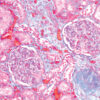
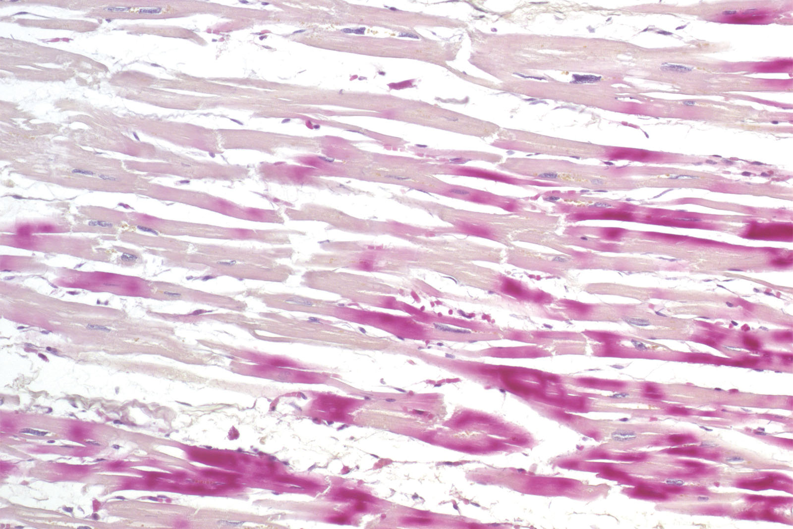
Histological diagnosis of ischemia in the early phase of myocardial infarction using the standard hematoxylin-eosin histological methods and a light microscope is exceptionally delicate. The reason for that is minimal histopathological changes occurring on the cardiac muscle during the first six hours of symptoms. However, staining the section using the kit consisting of hematoxylin, basic fuchsin and picric acid enables a histological overview of early changes on the cardiac muscle caused by ischemia or myocardial infarction.
H.B.F.P. kit
Three-reagent Hematoxylin-Basic Fuchsin-Picric acid staining kit for detection of cardiac muscle changes after ischemia or myocardial infarction. H.B.F.P. kit is a non-enzymatic histochemical technique for detection of early myocardial ischemia with vivid contrast.
Description
Additional information
| Size | |
|---|---|
| Brand | |
| Stain pack | |
| Stain Category | Muscle and Connective Tissue |
Downloads
Related products

Stain Kits
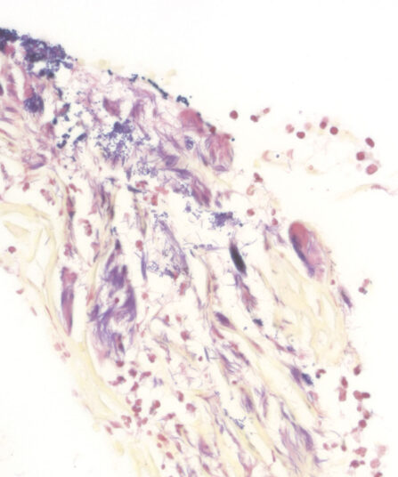
TB-Stain Histo kit
Three-reagent kit for staining acid-fast bacteria (pathogenic mycobacteria) in histology sections, sputum, smears and culture smears according to Ziehl-Neelsen. Heating of the carbol-fuchsin solution is avoided in this protocol hence omitting the release of hazardous phenolic vapours.
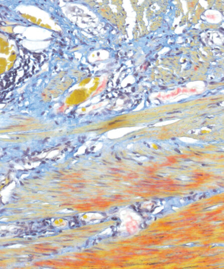
Martius Scarlet Blue (MSB) kit, 6x100ml+1x250ml
Seven-reagent kit used for fibrin visualisation, especially of older clusters. This method is a modification of Masson Trichrome and is ideal for studying connective tissue and vascular pathology.

Stains Reagents And Dyes
Stain Kits
BioGram Histo kit
Five-reagent kit for identification of bacteria according to Gram. For differentiation between Gram-positive and Gram-negative bacteria in histology sections.
Stain Kits
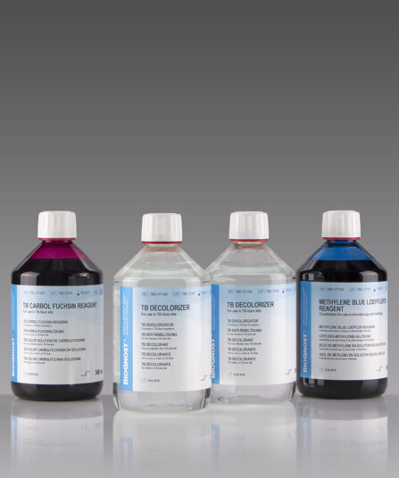
TB-Stain Hot Kit
Three-reagent kit for staining acid-fast bacteria. Contains TB Carbol Fuchsin reagent, double amount of TB Decolorizer and Methylene Blue Loeffler’s reagent as counterstain.
4 x 100ml bottles.
Stain Kits
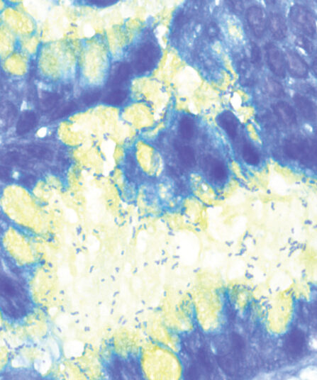
Alcian Yellow Toluidine Blue kit
Six-reagent kit for staining Helicobacter pylori in gastric tissue sections. This method is one of the most popular non-silver methods for staining of H. pylori, where bacteria are stained blue in contrast to yellow mucins.
Stain Kits
Martius Scarlet Blue (MSB) kit
Seven-reagent kit used for fibrin visualisation, especially of older clusters. This method is a modification of Masson Trichrome and is ideal for studying connective tissue and vascular pathology.
Stain Kits
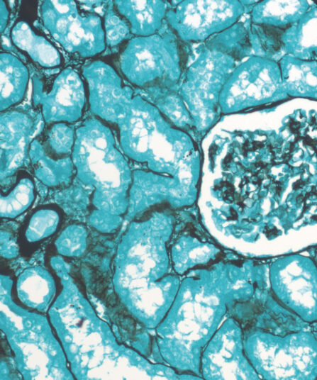
Grocott kit, stabilised
Seven-reagent kit for visualization of fungi and histological argentaffin structures in general (such as basal membranes). Green counterstain provides clear and visually rich contrast to target structures stained black.