Stain Kits
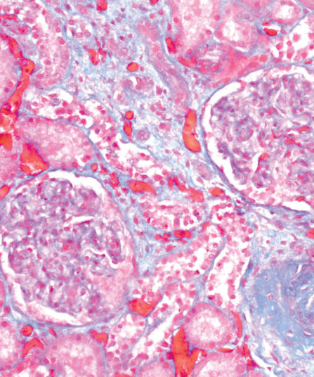
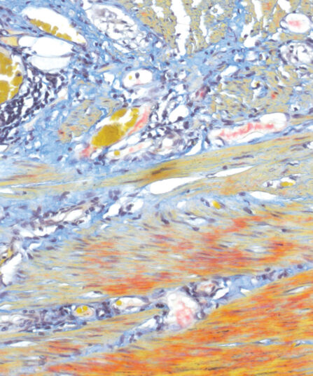
A.F.O.G. kit
Six-reagent Acid Fuchsin Orange G kit for selective staining of glomerular protein deposits and collagen in kidney biopsies. Nuclear stain is obtained with Weigert ferric hematoxylin, cytoplasm with Orange G and highly selective collagen stain with Aniline blue.
Stain Kits
Azan Trichrome kit
Five-reagent kit for connective tissue staining according to Mallory. Used for visualisation of muscle fibres, collagen, glial cells, glomerular cells and erythrocytes.
Stain Kits
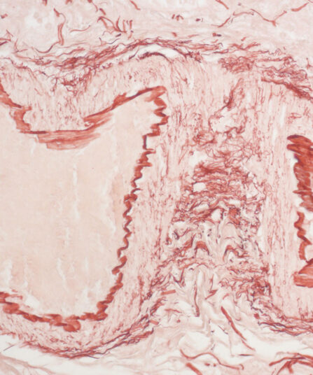
Elastica-Van Gieson kit
Four-reagent kit for staining elastic fibres and differentiation between elastic tissue, collagen and other types of connective tissue. The rapid method enables a satisfactory result with shorter section staining time.
Stain Kits
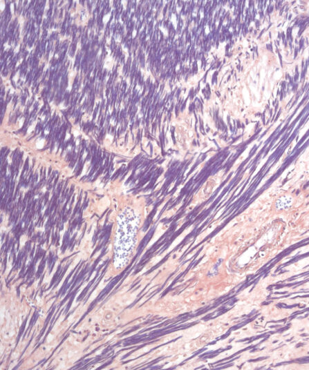
Gomori Trichrome kit
Five-reagent kit for staining muscle, collagen fibre and nuclei, contains blue counterstain. The kit can be used to contrast skeletal, cardiac or smooth muscle.
Stain Kits
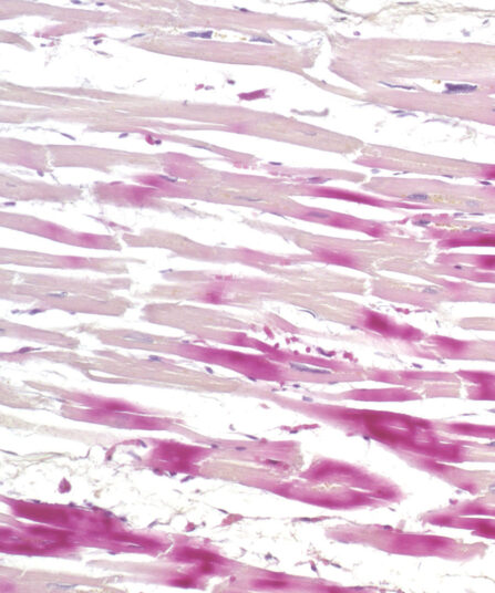
H.B.F.P. kit
Three-reagent Hematoxylin-Basic Fuchsin-Picric acid staining kit for detection of cardiac muscle changes after ischemia or myocardial infarction. H.B.F.P. kit is a non-enzymatic histochemical technique for detection of early myocardial ischemia with vivid contrast.
Stain Kits
Hematoxylin P.T.A. kit
Four-reagent Hematoxylin-Phosphotungstic Acid kit for differentiation of smooth and striated muscle tissues as well as for the detection of fibrin, collagen and elements of the central nervous system according to Mallory.
Stain Kits
Hematoxylin W kit
Acid-resistant hematoxylin according to Weigert. Two-reagent kit that stains the nuclei blue-black, often a component of special staining kits for connective tissues.
Stain Kits
Hematoxylin W kit 2x500ml
Acid-resistant hematoxylin according to Weigert. Two-reagent kit that stains the nuclei blue-black, often a component of special staining kits for connective tissues.
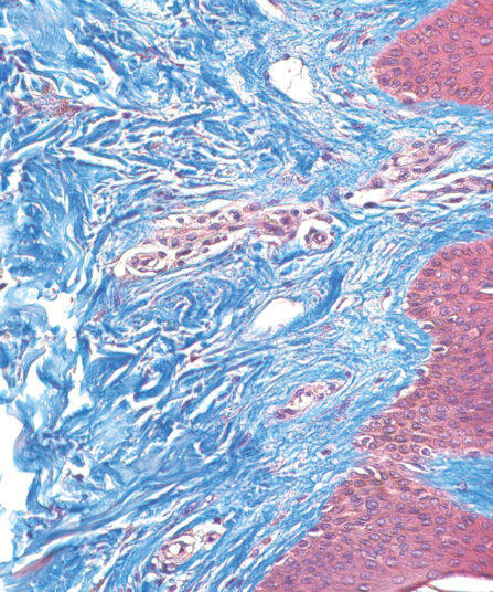
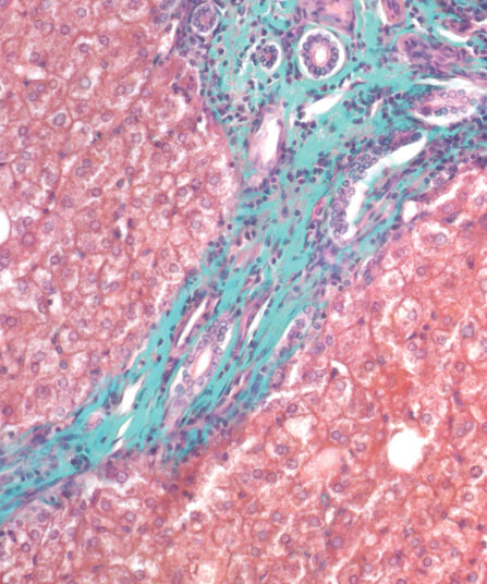
Stain Kits
Mallory Trichrome kit
Three-reagent staining kit for connective tissue visualisation and detection of collagen, cartilage, muscle, elastic fibres, mucous, pituarity cells, reticulum, bones, amyloid and erythrocytes.
Stain Kits
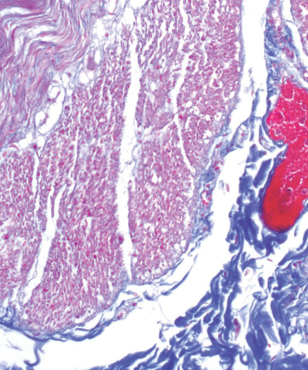
Martius Scarlet Blue (MSB) kit
Seven-reagent kit used for fibrin visualisation, especially of older clusters. This method is a modification of Masson Trichrome and is ideal for studying connective tissue and vascular pathology.
Martius Scarlet Blue (MSB) kit, 6x100ml+1x250ml
Seven-reagent kit used for fibrin visualisation, especially of older clusters. This method is a modification of Masson Trichrome and is ideal for studying connective tissue and vascular pathology.
Stain Kits
Masson Trichrome kit
Seven-reagent kit for staining muscle and collagen fibers with a blue counterstain. It is also used for visualizing gametes, nuclei, neurofibrils, glial cells, keratins and intercellular fibrils. The kit may be useful for detecting collagen in smooth muscle cancer or diseases like cirrhosis.
Stain Kits
Masson-Goldner Trichrome kit
Seven-reagent kit for staining muscle and collagen fibres with green counterstain. It is also used for visualising gametes, nuclei, neurofibrils, glial cells, keratins, intercellular fibrils and for differentiation of smooth muscle fibres and collagens.
Stain Kits
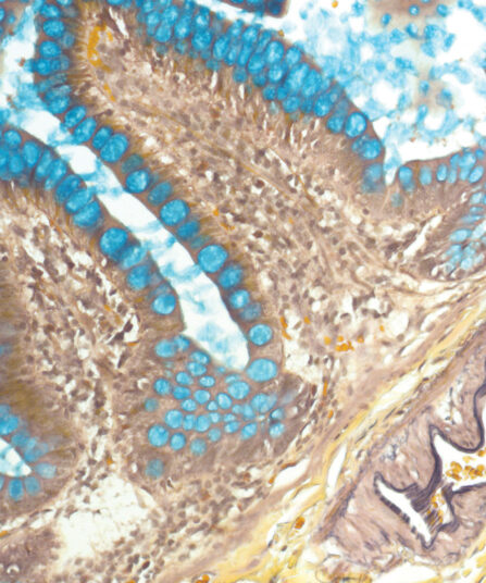
Orcein kit
Five-reagent kit for visualisation of hepatitis B surface antigen (HBsAg) seen as viral inclusion bodies in hepatocytes, for elastic fibres and copper associated protein in tissue sections. It can be used with frozen sections.
Stain Kits
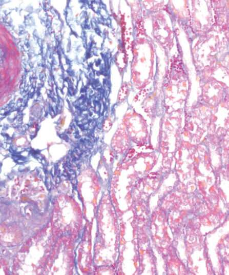
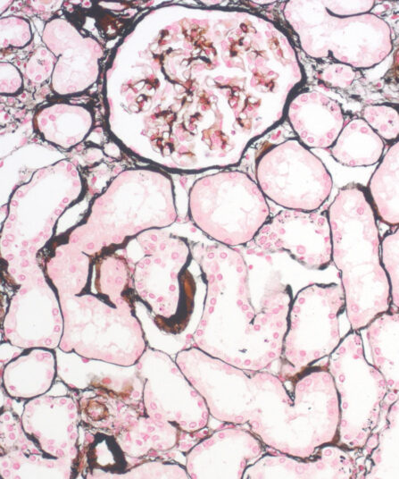
P.A.S.M. / Jones kit, stabilised
Seven-reagent Periodic Acid Silver Methenamine kit for staining kidney glomerular basement membranes. Kit includes red counterstain which provides visually rich contrast to target structures stained black.
Stain Kits
Paraldehyde Fuchsin kit
Seven-reagent kit according to Gomori for detecting pathological changes in elastic fibres. It also stains mast cell granules, beta granules in pancreatic islets, neurosecretory material, mast cell granules and beta cells in the pituitary gland.
Stain Kits
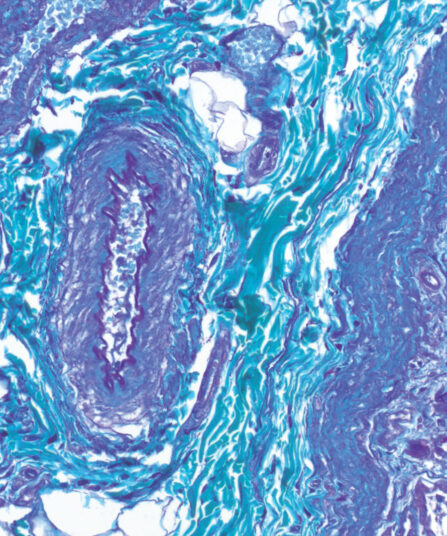
Reticulin Contrast kit
Nine-reagent kit for detecting argyrophilic reticulin fibres according to Gordon and Sweets. The kit contains gold chloride solution that enhances visualisation of reticulin fibres and it also contains Nuclear Fast Red (Kernechtrot) reagent that enables fine contrasting background.
Stain Kits
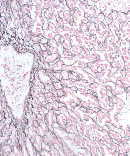
Reticulin kit
Seven-reagent kit for detecting argyrophilic reticulin fibres. It clearly differentiates between collagen and reticulin and nerve fibres and connective tissue. The main function of reticular fibres is to provide support and are normally found in liver, lymph nodes, spleen and kidneys.
Stain Kits
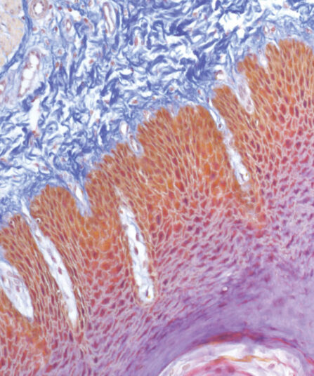
Van Gieson Trichrome kit
Three-reagent kit for staining collagen fibres, muscle tissue, keratinized epithelium, cytoplasm, glial fibres and erythrocytes. Used for differentiation between collagen and smooth fibres in tumours and various other diseases.
Stain Kits
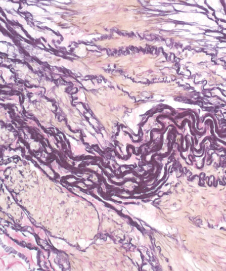
Verhoeff kit
Six-reagent kit for detecting atrophy of elastic tissue in cases of emphysema, thinning and loss of elastic fibres in arteriosclerosis and other vascular diseases, or whether blood vessels have been invaded by a tumour.