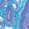
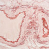
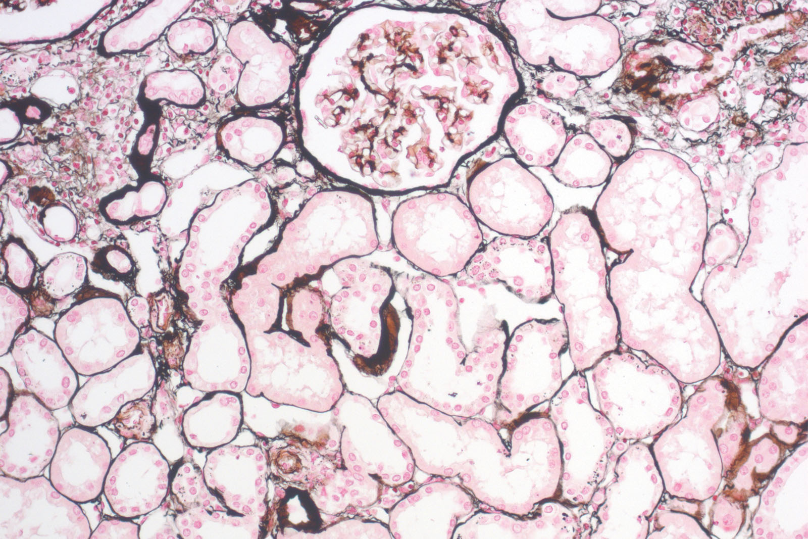
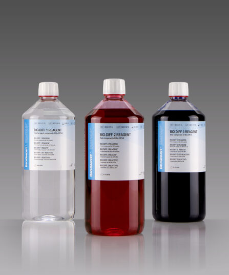
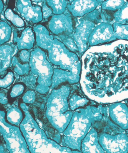
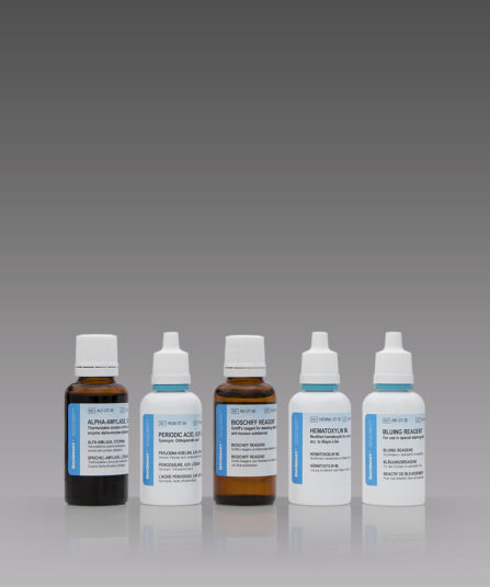
P.A.S.M. / Jones kit is used in histology for visualizing argentaffin structures, especially kidney membranes, but also fungi and certain pathogen organisms. Staining procedure starts with periodic acid solution being used to oxidize 1,2-glycols to aldehydes. During incubation in silver-methenamine-borate working solution aldehydes are reduced and at the same time cause reduction of silver ions to metallic silver that manifests as brown to black structures on the section. This is followed by toning the solution with gold chloride solution that additionally improves staining intensity of target structures (fungi, basal membranes and others), and it reduces background staining. Excessive unbound silver-gold bonds is removed by rinsing the section with sodium thiosulfate solution. Finally, the sections are exposed to Nuclear Fast Red (Kernechtrot) counterstain that stains background structures red; that in turn creates clear and visually rich contrast to target structures (coloured in brown-black).
This is new and improved formulation for P.A.S.M./Jones staining. Kit is stable at room temperature, do not store at lower temperature!
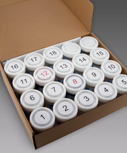
P.A.S.M. / Jones kit, stabilised
Seven-reagent Periodic Acid Silver Methenamine kit for staining kidney glomerular basement membranes. Kit includes red counterstain which provides visually rich contrast to target structures stained black.
Description
Additional information
| Size | |
|---|---|
| Brand | |
| Stain pack | |
| Stain Category | Muscle and Connective Tissue |
Downloads
Related products

Stain Kits
TB-Stain Histo kit
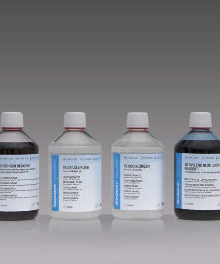
Three-reagent kit for staining acid-fast bacteria (pathogenic mycobacteria) in histology sections, sputum, smears and culture smears according to Ziehl-Neelsen. Heating of the carbol-fuchsin solution is avoided in this protocol hence omitting the release of hazardous phenolic vapours.
Stain Kits
BioGram Histo kit
Five-reagent kit for identification of bacteria according to Gram. For differentiation between Gram-positive and Gram-negative bacteria in histology sections.
Stain Kits
Bio-Diff Kit 3 X 1L
Three-reagent kit that contains fixative agent, red and blue components for fast and effective staining. Each kit contains buffer tablets for consistent staining results.
3×100 ml bottles
Stain Kits
Grocott kit, stabilised
Seven-reagent kit for visualization of fungi and histological argentaffin structures in general (such as basal membranes). Green counterstain provides clear and visually rich contrast to target structures stained black.
Stain Kits
TB-Stain ECO Kit
Three-reagent phenol-free kit for staining acid-fast bacteria. Contains TB-Fuchsin reagent, double amount of TB Decolorizer and Methylene Blue Loeffler’s reagent as counterstain.
5 x 100ml bottles.
Stain Kits
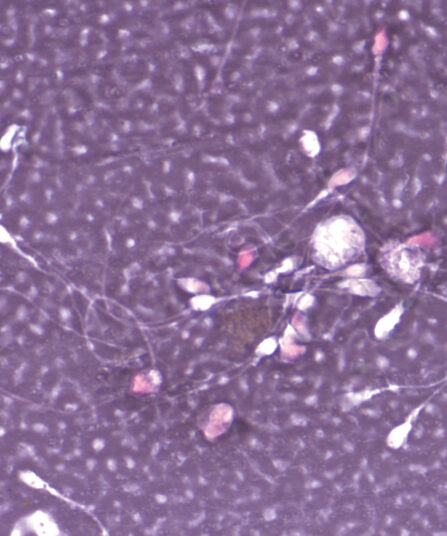
Eosin-Nigrosin Vital
Fast detection (one-step detection) of sperm vitality and visualisation of dead and living sperm cells with one reagent. A simple, easy and fast method for semen analysis.
HE Rapid Staining kit- frozen and paraffin sections
Ready-to-use eight-reagent kit (in 16 containers that can be used as staining jars) for rapid HE staining of frozen and paraffin tissue sections in histopathology. Contains xylene substitute as clearing agent and xylene substitute-based medium for permanent section covering.
For 100 tests.
Stain Kits
P.A.S. Diastase Kit
BioGnost’s P.A.S. Diastase kit is most commonly used for identifying glycogen in liver. Periodic acid enables the molecules containing glycol groups to create aldehydes affected by Schiff’s reagent staining them violet (magenta). Specific stains are created by applying the PAS method on unsubsti-tuted polysaccharides, mucoproteins and glycoproteins, glycolipids and phospholipids. Alpha-amylase enzyme (also known as diastasis) is used for differentiation between glycogen and other PAS-positive structures by dissolving 1→4 glycosidic bonds, causing the glycogen to remain unstained after the PAS reaction. BioGnost’s P.A.S. Diastase kit uses thermostable enzyme which does not require heating to +37°C to be active, but incubat-ing the section at +37°C is preferred in order to achieve better glycogen breakdown. The same tissue section is used as negative control for this reaction, but the sample is not treated using alpha-amylase.
For 100 tests.