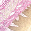

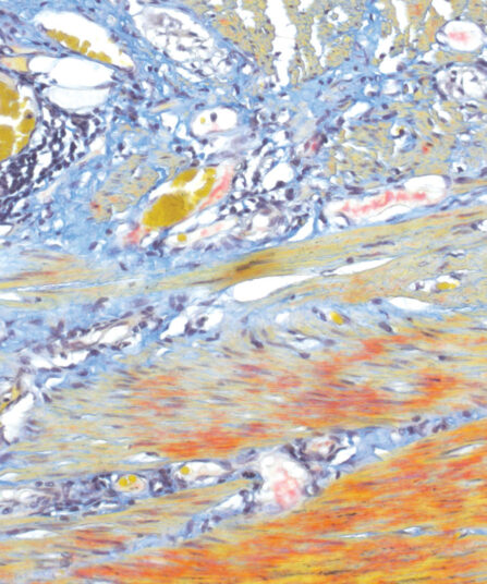
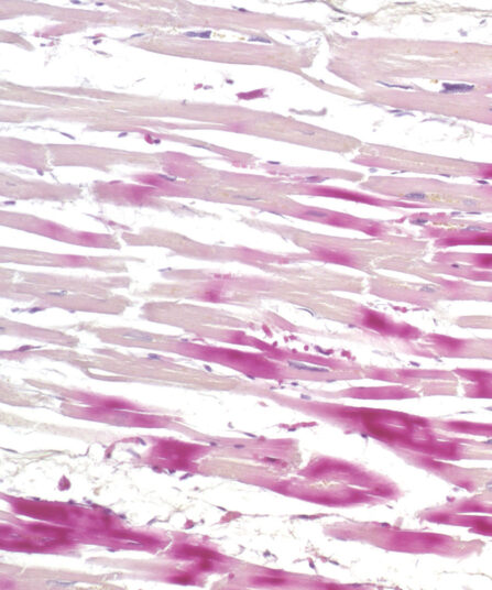
Reticulin Contrast kit is used for identification and easier visualisation of argentaffin reticular fibres in connective tissue. Reticulin provides structural support. It is found in the liver, spleen and kidneys. Reticulin fibres are clearly defined in the healthy liver; necrotic and cirrhotic liver has discontinuous fibres. The visualisation is based on silver depositions on reticulin fibres. The tissue sample must be oxidized with potassium permanganate. Silver is formed from ammonia solution containing silver nitrate and is deposited in the form of brown sediment on reticulin fibres. Formalin acts as a reducing agent and accelerates the procedure. Unbound silver is washed away using sodium thiosulfate. Reticulin Contrast kit also contains a gold chloride solution that stabilises and tones the section’s image. The kit contains Nuclear Fast Red (Kernechtrot) counterstain.
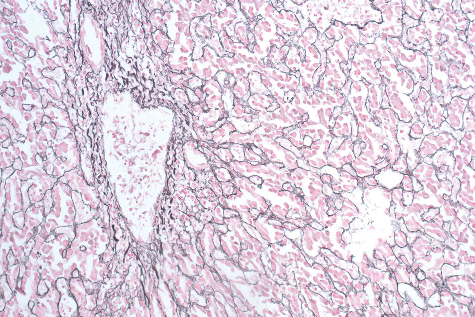
Reticulin Contrast kit
Nine-reagent kit for detecting argyrophilic reticulin fibres according to Gordon and Sweets. The kit contains gold chloride solution that enhances visualisation of reticulin fibres and it also contains Nuclear Fast Red (Kernechtrot) reagent that enables fine contrasting background.
Description
Additional information
| Size | |
|---|---|
| Brand | |
| Stain pack | |
| Stain Category | Muscle and Connective Tissue |
Downloads
Related products
Stain Kits
Giemsa HP kit
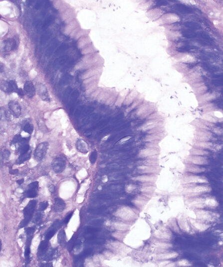
Four-reagent kit for staining Helicobacter pylori in gastroscopic sections according to Lennart. Advantages of this method for detecting H. pylori are sensitive and reproducible results and easy performance.
Stain Kits
BioGram Histo kit
Five-reagent kit for identification of bacteria according to Gram. For differentiation between Gram-positive and Gram-negative bacteria in histology sections.
Stain Kits
Martius Scarlet Blue (MSB) kit
Seven-reagent kit used for fibrin visualisation, especially of older clusters. This method is a modification of Masson Trichrome and is ideal for studying connective tissue and vascular pathology.
Stain Kits
Elastica-Van Gieson kit
Four-reagent kit for staining elastic fibres and differentiation between elastic tissue, collagen and other types of connective tissue. The rapid method enables a satisfactory result with shorter section staining time.

Stain Kits
TB-Stain Fluorescent Kit
Three-reagent kit for fluorescence-microscopic detection of acid-fast bacteria. Contains TB Auramine-Rhodamine reagent, double amount of TB Decolorizer Fluorescent and TB Permanganate reagent as counterstain.
4 x 100ml bottles.
Stain Kits
P.A.S. Diastase Kit
BioGnost’s P.A.S. Diastase kit is most commonly used for identifying glycogen in liver. Periodic acid enables the molecules containing glycol groups to create aldehydes affected by Schiff’s reagent staining them violet (magenta). Specific stains are created by applying the PAS method on unsubsti-tuted polysaccharides, mucoproteins and glycoproteins, glycolipids and phospholipids. Alpha-amylase enzyme (also known as diastasis) is used for differentiation between glycogen and other PAS-positive structures by dissolving 1→4 glycosidic bonds, causing the glycogen to remain unstained after the PAS reaction. BioGnost’s P.A.S. Diastase kit uses thermostable enzyme which does not require heating to +37°C to be active, but incubat-ing the section at +37°C is preferred in order to achieve better glycogen breakdown. The same tissue section is used as negative control for this reaction, but the sample is not treated using alpha-amylase.
For 100 tests.
Stain Kits
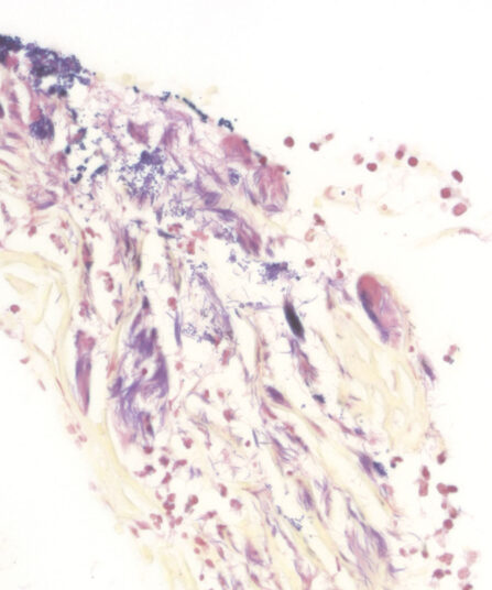
Warthin Starry kit
Five-reagent kit for staining Spirochaeta, Helicobacter pylori, Microsporidia and Legionella pneumophila. The kit contains 12 jars with gelatine that enables both incubation and staining of sections, as well as other reagents that enable precipitation of silver on the bacterial surface. The bacteria are found in the mucus of the surface epithelium, in the apical gastric glands and in the gastric mucosa.
Stain Kits
H.B.F.P. kit
Three-reagent Hematoxylin-Basic Fuchsin-Picric acid staining kit for detection of cardiac muscle changes after ischemia or myocardial infarction. H.B.F.P. kit is a non-enzymatic histochemical technique for detection of early myocardial ischemia with vivid contrast.