
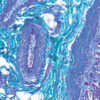
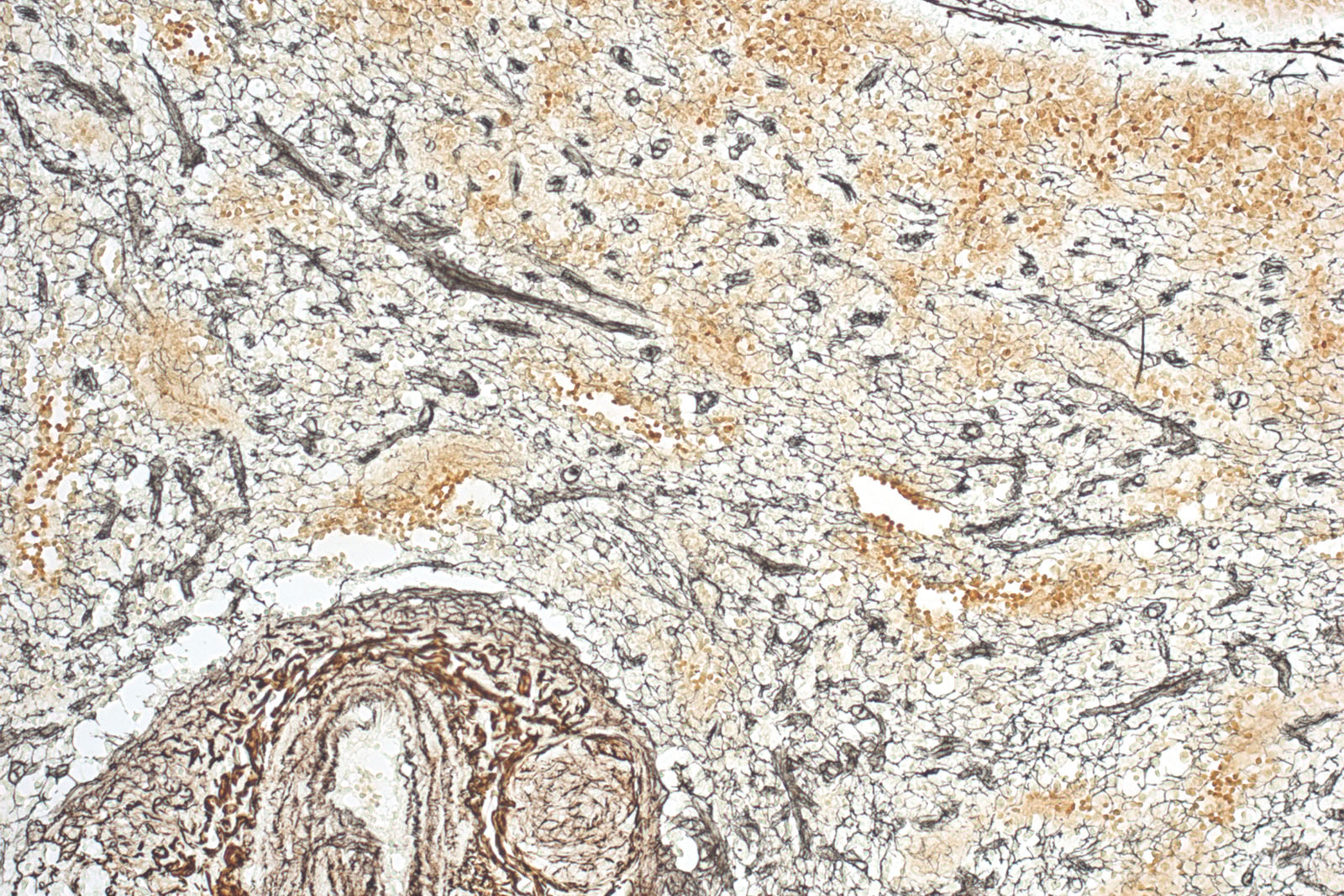
Reticulin kit is used for identification and easier visualisation of argentaffin reticular fibres in connective tissue. Reticulin provides structural support. It is found in the liver, spleen and kidneys. Reticulin fibres are clearly defined in the healthy liver; necrotic and cirrhotic liver has discontinuous fibres. The visualisation is based on silver depositions on reticulin fibres. The tissue sample must be oxidized with potassium permanganate. Silver is formed from ammonia solution containing silver nitrate and is deposited in the form of brown sediment on reticulin fibres. Formalin acts as a reducing agent and accelerates the procedure. Unbound silver is washed away using sodium thiosulfate.
Reticulin kit
Seven-reagent kit for detecting argyrophilic reticulin fibres. It clearly differentiates between collagen and reticulin and nerve fibres and connective tissue. The main function of reticular fibres is to provide support and are normally found in liver, lymph nodes, spleen and kidneys.
Description
Additional information
| Size | |
|---|---|
| Brand | |
| Stain pack | |
| Stain Category | Muscle and Connective Tissue |
Downloads
Related products
Stain Kits
TB-Stain ECO Kit
Three-reagent phenol-free kit for staining acid-fast bacteria. Contains TB-Fuchsin reagent, double amount of TB Decolorizer and Methylene Blue Loeffler’s reagent as counterstain.
5 x 100ml bottles.
Stain Kits
TB-Stain Auramine O Kit
Three-reagent kit for staining acid-fast bacteria using fluorescence method. Contains TB Auramine O reagent, double amount of TB Decolorizer Fluorescent and counterstain of TB Permanganate reagent.
4 x 100ml bottles.
Stain Kits
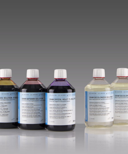
BioGram 4 kit
Four-reagent kit for identification of bacteria according to Gram. Kit contains Gram Crystal Violet 1% solution, stabilized Gram Lugol solution, double amount of Gram Decolorizer solution 2 and Gram Safranin solution as counterstain.
5×100 ml bottles
Stain Kits
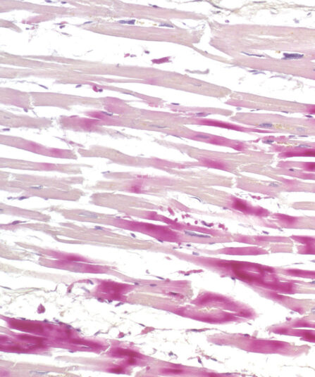
H.B.F.P. kit
Three-reagent Hematoxylin-Basic Fuchsin-Picric acid staining kit for detection of cardiac muscle changes after ischemia or myocardial infarction. H.B.F.P. kit is a non-enzymatic histochemical technique for detection of early myocardial ischemia with vivid contrast.

Stains Reagents And Dyes

Stain Kits
Bio-Diff kit 3 x 500ml
Three-reagent kit that contains fi xative agent, red and blue components for fast and effective staining. Each kit contains buffer tablets for consistent staining results.
Stain Kits
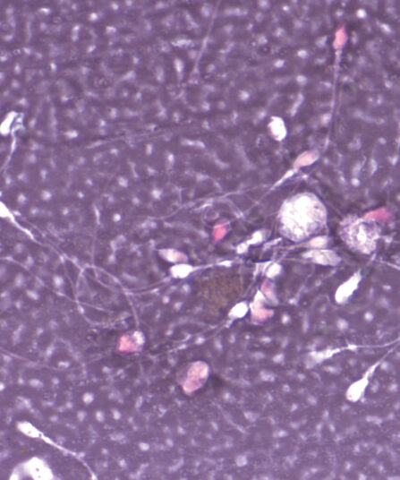
Eosin-Nigrosin Vital
Fast detection (one-step detection) of sperm vitality and visualisation of dead and living sperm cells with one reagent. A simple, easy and fast method for semen analysis.
Stain Kits
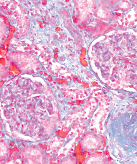
Gomori Trichrome kit
Five-reagent kit for staining muscle, collagen fibre and nuclei, contains blue counterstain. The kit can be used to contrast skeletal, cardiac or smooth muscle.