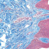
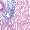
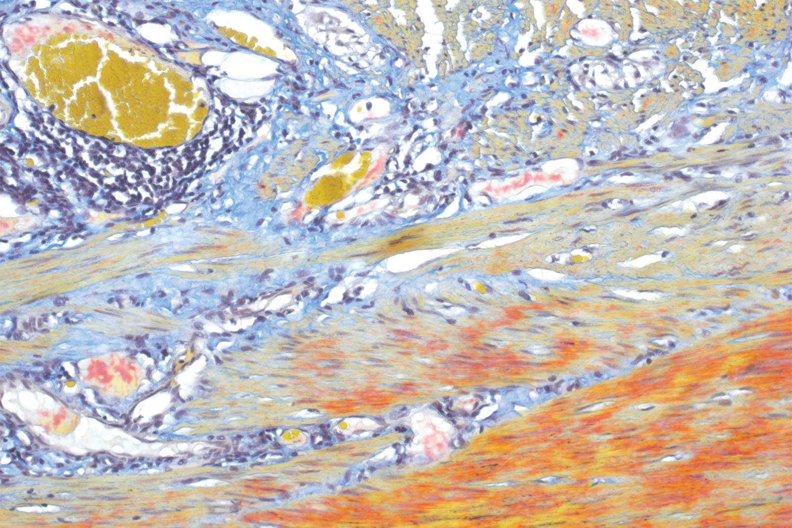
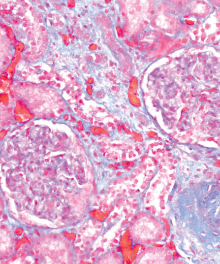
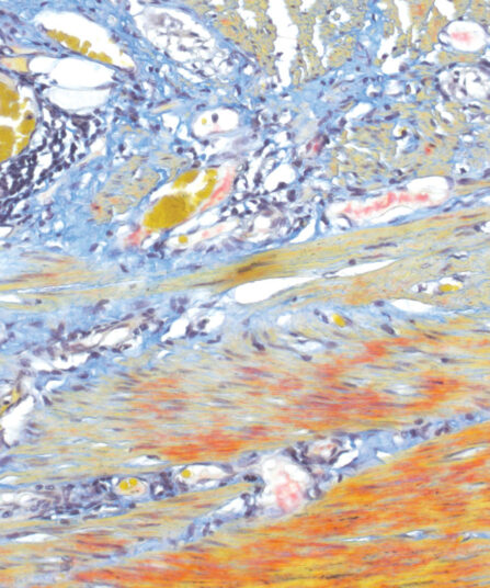
Histology, cytology and other related scientific disciplines study the microscopic anatomy of tissues and cells. In order to demonstrate a good tissue and cellular structure, the samples need to be stained in a correct manner. Martius Scarlet Blue (MSB) staining technique is used for fibrin visualisation, especially of older clusters. This method is a modified Masson Trichrome method and it is ideal for studying connective tissue and vascular pathology.
Martius Scarlet Blue (MSB) kit
Seven-reagent kit used for fibrin visualisation, especially of older clusters. This method is a modification of Masson Trichrome and is ideal for studying connective tissue and vascular pathology.
Description
Additional information
| Size | |
|---|---|
| Brand | |
| Stain pack | |
| Stain Category | Muscle and Connective Tissue |
Downloads
Related products
Stain Kits
Alcian Yellow Toluidine Blue kit
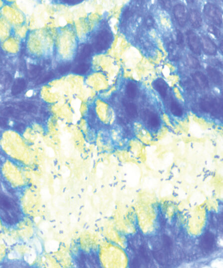
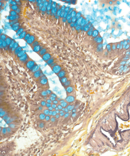
Six-reagent kit for staining Helicobacter pylori in gastric tissue sections. This method is one of the most popular non-silver methods for staining of H. pylori, where bacteria are stained blue in contrast to yellow mucins.
Stain Kits
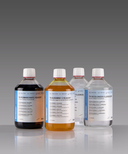
TB-Stain Auramine O Kit
Three-reagent kit for staining acid-fast bacteria using fluorescence method. Contains TB Auramine O reagent, double amount of TB Decolorizer Fluorescent and counterstain of TB Permanganate reagent.
4 x 100ml bottles.
Stain Kits
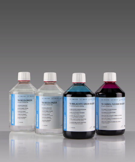
TB-Stain Cold Kit
Three-reagent kit for staining acid-fast bacteria according to Kinyoun. Contains TB Carbol Fuchsin reagent, double amount of TB Decolorizer and TB Malachite Green reagent as counterstain.
4 x 100ml bottles.
Stain Kits
Warthin Starry kit
Five-reagent kit for staining Spirochaeta, Helicobacter pylori, Microsporidia and Legionella pneumophila. The kit contains 12 jars with gelatine that enables both incubation and staining of sections, as well as other reagents that enable precipitation of silver on the bacterial surface. The bacteria are found in the mucus of the surface epithelium, in the apical gastric glands and in the gastric mucosa.
Stain Kits
Gomori Trichrome kit
Five-reagent kit for staining muscle, collagen fibre and nuclei, contains blue counterstain. The kit can be used to contrast skeletal, cardiac or smooth muscle.

Stain Kits
TB-Stain Histo kit
Three-reagent kit for staining acid-fast bacteria (pathogenic mycobacteria) in histology sections, sputum, smears and culture smears according to Ziehl-Neelsen. Heating of the carbol-fuchsin solution is avoided in this protocol hence omitting the release of hazardous phenolic vapours.
Martius Scarlet Blue (MSB) kit, 6x100ml+1x250ml
Seven-reagent kit used for fibrin visualisation, especially of older clusters. This method is a modification of Masson Trichrome and is ideal for studying connective tissue and vascular pathology.