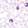
Gram staining is a method of differentiating bacterial species and it is commonly known and used in microbiology. It is also one of the most frequently used diagnostic methods in hospital and clinical laboratories. Gram staining differentiates bacteria into two groups: Gram-positive and Gram-negative. That division is based on the two groups’ bacterial membrane structural differences, i.e. their capability of retaining the dye. Gram-positive bacteria have a thicker cellular membrane which enables retaining the dye inside the cell by treating them with iodine solution that creates insoluble iodine and primary dye complex. Gram-negative bacteria have thinner cellular membrane structure which cannot retain the dye. It washes away through the membrane, and using counterstaining forms the basis for differentiating between the two bacteria groups. BioGnost’s BioGram Histo kit contains Gram Crystal Violet 1% solution, stabilised Gram Lugol solution, two packages of Gram Decolouriser 2 solution, Gram Safranin solution and two packages of picric acid in acetone. Its characteristics make it an optimal bacteria staining agent which provides consistent results.
BioGram Histo kit
Five-reagent kit for identification of bacteria according to Gram. For differentiation between Gram-positive and Gram-negative bacteria in histology sections.
Description
Additional information
| Size | |
|---|---|
| Brand | |
| Stain pack | |
| Stain Category | Fungi, Bacteria and Parasites |
Downloads
Related products
Stain Kits
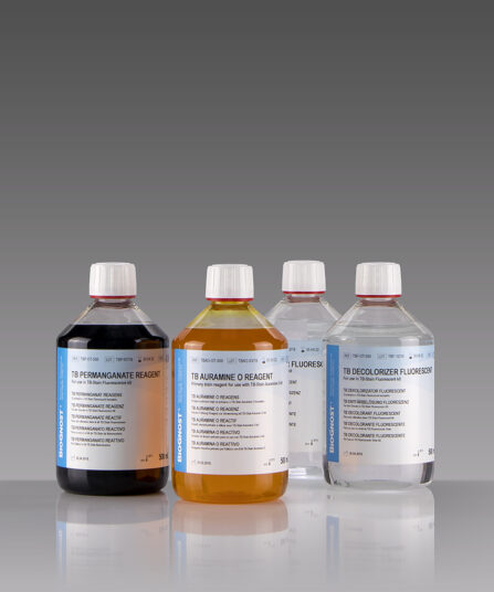
TB-Stain Auramine O Kit
Three-reagent kit for staining acid-fast bacteria using fluorescence method. Contains TB Auramine O reagent, double amount of TB Decolorizer Fluorescent and counterstain of TB Permanganate reagent.
4 x 100ml bottles.
Stain Kits
TB-Stain Cold Kit
Three-reagent kit for staining acid-fast bacteria according to Kinyoun. Contains TB Carbol Fuchsin reagent, double amount of TB Decolorizer and TB Malachite Green reagent as counterstain.
4 x 100ml bottles.
Stain Kits
Field kit 500ml
Ready to use two-reagent kit for rapid and efficient staining and detection of parasites in haematology samples. Primarily used for staining thin and thick blood smears (dense drop) for purpose of diagnosing blood parasites. Reagents are stored in containers that can be used as staining jars.
Stain Kits
Gomori Trichrome kit
Five-reagent kit for staining muscle, collagen fibre and nuclei, contains blue counterstain. The kit can be used to contrast skeletal, cardiac or smooth muscle.

Stain Kits
TB-Stain Fluorescent Kit
Three-reagent kit for fluorescence-microscopic detection of acid-fast bacteria. Contains TB Auramine-Rhodamine reagent, double amount of TB Decolorizer Fluorescent and TB Permanganate reagent as counterstain.
4 x 100ml bottles.
Stain Kits
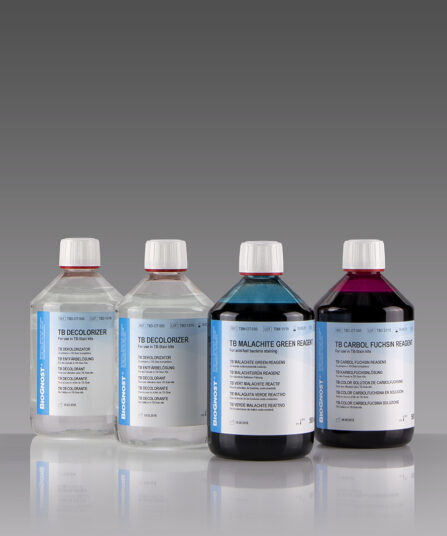
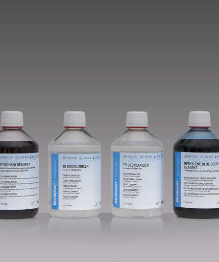
TB-Stain Hot Kit
Three-reagent kit for staining acid-fast bacteria. Contains TB Carbol Fuchsin reagent, double amount of TB Decolorizer and Methylene Blue Loeffler’s reagent as counterstain.
4 x 100ml bottles.
Stain Kits
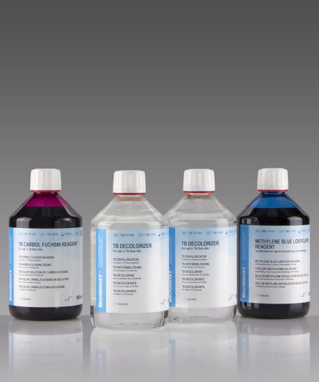
TB-Stain ECO Kit
Three-reagent phenol-free kit for staining acid-fast bacteria. Contains TB-Fuchsin reagent, double amount of TB Decolorizer and Methylene Blue Loeffler’s reagent as counterstain.
5 x 100ml bottles.
Stain Kits
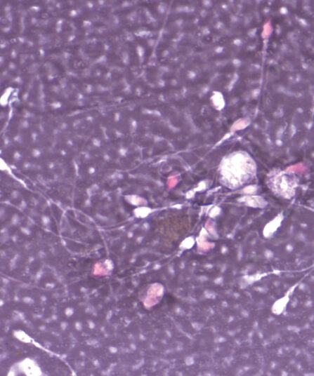
Eosin-Nigrosin Vital
Fast detection (one-step detection) of sperm vitality and visualisation of dead and living sperm cells with one reagent. A simple, easy and fast method for semen analysis.