Stain Kits
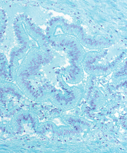
Feulgen kit
Five-reagent DNA staining kit according to Feulgen. For use in semiquantitative DNA determination in histological and cytological samples. The specimen is first treated with hydrochloric acid creating an aldehyde group of DNA that can be visualised by Schiff (BioSchiff) reagent. This reaction is specific for nuclear DNA.
Stain Kits
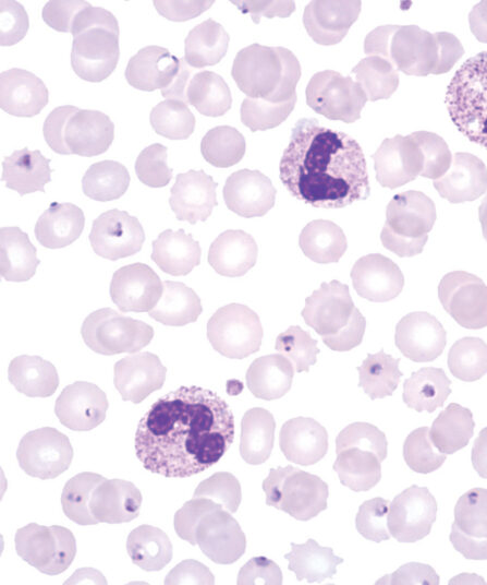
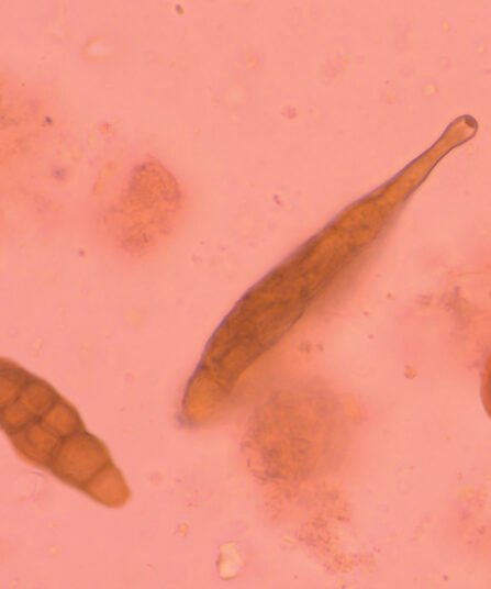
Field kit
Ready to use two-reagent kit for rapid and efficient staining and detection of parasites in haematology samples. Primarily used for staining thin and thick blood smears (dense drop) for purpose of diagnosing blood parasites. Reagents are stored in containers that can be used as staining jars.
Stain Kits
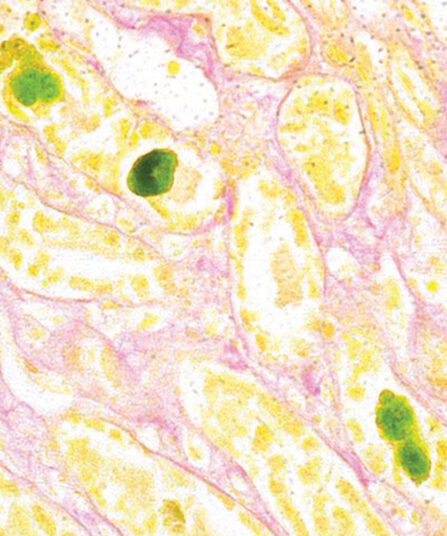
Fouchet-Van Gieson kit
Three-reagent kit for visualisation of bilirubin and collagen according to Kutllick. Bilirubin is a yellow-brown pigment, but changes to green due to oxidation induced by Fouchet reaction. Green bilirubin can easily be detected on yellowish and pink coloured background.
Stain Kits
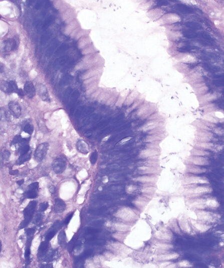
Giemsa HP kit
Four-reagent kit for staining Helicobacter pylori in gastroscopic sections according to Lennart. Advantages of this method for detecting H. pylori are sensitive and reproducible results and easy performance.
Stain Kits
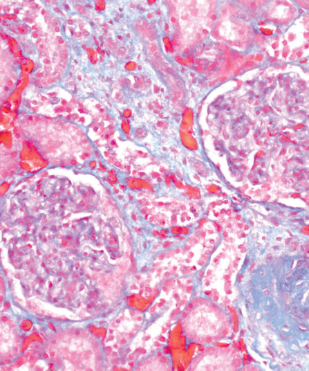
Gomori Trichrome kit
Five-reagent kit for staining muscle, collagen fibre and nuclei, contains blue counterstain. The kit can be used to contrast skeletal, cardiac or smooth muscle.
Stain Kits
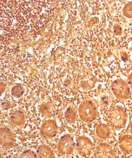
Grimelius kit
Five-reagent kit for staining argyrophilic granules. Grimelius kit can be used for the detection of secretory intracytoplasmatic granules specific for carcinoid tumours and for identification of neuroendocrine cells.
Stain Kits
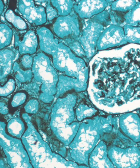
Grocott kit, stabilised
Seven-reagent kit for visualization of fungi and histological argentaffin structures in general (such as basal membranes). Green counterstain provides clear and visually rich contrast to target structures stained black.
Stain Kits
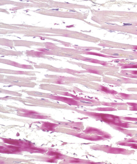
H.B.F.P. kit
Three-reagent Hematoxylin-Basic Fuchsin-Picric acid staining kit for detection of cardiac muscle changes after ischemia or myocardial infarction. H.B.F.P. kit is a non-enzymatic histochemical technique for detection of early myocardial ischemia with vivid contrast.
HE Rapid Staining kit- frozen and paraffin sections
Ready-to-use eight-reagent kit (in 16 containers that can be used as staining jars) for rapid HE staining of frozen and paraffin tissue sections in histopathology. Contains xylene substitute as clearing agent and xylene substitute-based medium for permanent section covering.
For 100 tests.
Stain Kits
Hematoxylin P.T.A. kit
Four-reagent Hematoxylin-Phosphotungstic Acid kit for differentiation of smooth and striated muscle tissues as well as for the detection of fibrin, collagen and elements of the central nervous system according to Mallory.
Stain Kits
Hematoxylin W kit
Acid-resistant hematoxylin according to Weigert. Two-reagent kit that stains the nuclei blue-black, often a component of special staining kits for connective tissues.
Stain Kits
Hematoxylin W kit 2x500ml
Acid-resistant hematoxylin according to Weigert. Two-reagent kit that stains the nuclei blue-black, often a component of special staining kits for connective tissues.
Stain Kits
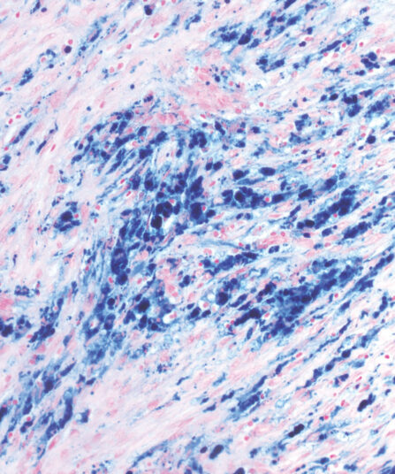
HemoGnost Perls kit
Three-reagent HemoGnost Perls (Prussian blue / Berlin blue) kit for the detection of reactive ferric (Fe3+) (not bound to haemoglobin) ions in cells. It is often applied on bone marrow and spleen cells.

Histanol 100 Denatured 100% Ethyl Alcohol
Histanol 100 is a high-quality histological clearing agent used in tissue processing and staining procedures. It offers excellent dewaxing and clearing performance with reduced odour, making it suitable for routine use in histopathology laboratories.
Stain Kits
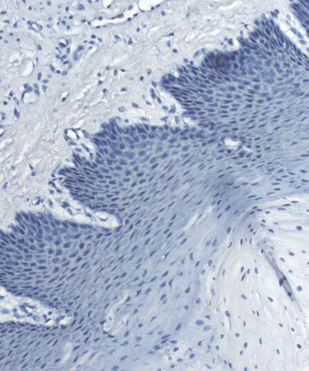
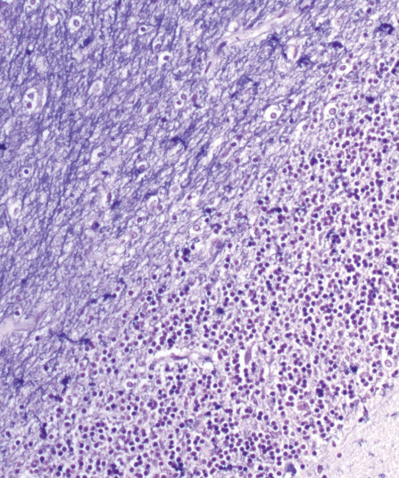
Luxol Fast Blue kit
Three-reagent kit for staining myelin and myelinated axons, Nissl bodies and phospholipids according to Kluwer-Barrera. The kit is used for identifying the basic neuronal structure in the brain or spinal cord sections.
Stain Kits
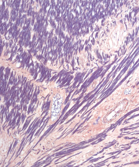
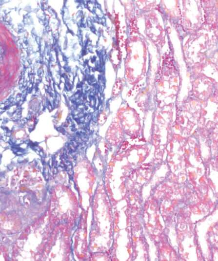
Mallory Trichrome kit
Three-reagent staining kit for connective tissue visualisation and detection of collagen, cartilage, muscle, elastic fibres, mucous, pituarity cells, reticulum, bones, amyloid and erythrocytes.
Stain Kits
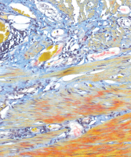
Martius Scarlet Blue (MSB) kit
Seven-reagent kit used for fibrin visualisation, especially of older clusters. This method is a modification of Masson Trichrome and is ideal for studying connective tissue and vascular pathology.
Martius Scarlet Blue (MSB) kit, 6x100ml+1x250ml
Seven-reagent kit used for fibrin visualisation, especially of older clusters. This method is a modification of Masson Trichrome and is ideal for studying connective tissue and vascular pathology.
Stain Kits
Masson Fontana kit
Six-reagent melanin and argentaffin granule staining kit, based on the reduction of silver nitrate to elemental silver. Melanin is a brown-black pigment normally present in the hair, skin, retina, iris and certain parts of CNS. Argentaffin granules are found in carcinoid tumours.