Stain Kits
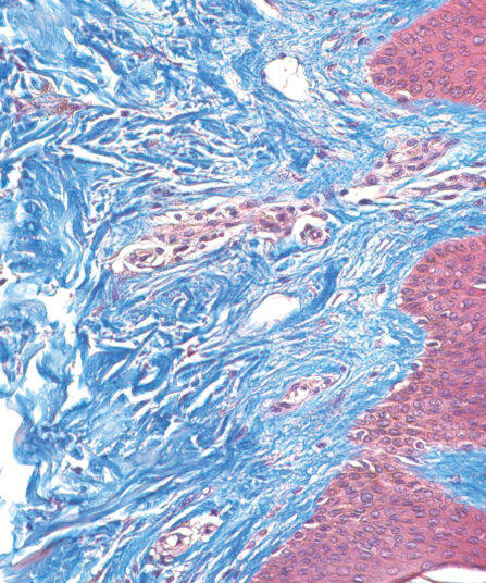
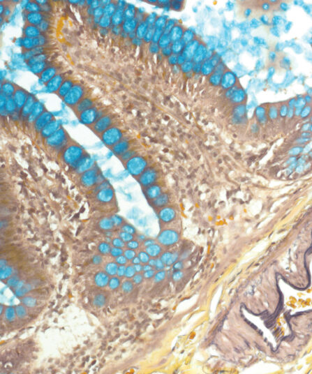
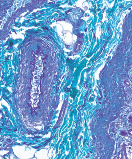
Masson Trichrome kit
Seven-reagent kit for staining muscle and collagen fibers with a blue counterstain. It is also used for visualizing gametes, nuclei, neurofibrils, glial cells, keratins and intercellular fibrils. The kit may be useful for detecting collagen in smooth muscle cancer or diseases like cirrhosis.
Stain Kits
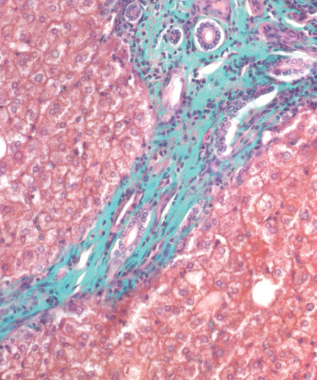
Masson-Goldner Trichrome kit
Seven-reagent kit for staining muscle and collagen fibres with green counterstain. It is also used for visualising gametes, nuclei, neurofibrils, glial cells, keratins, intercellular fibrils and for differentiation of smooth muscle fibres and collagens.
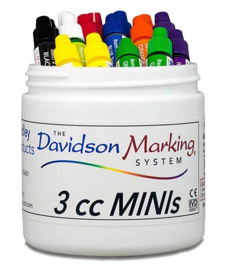
Mini Tissue Marking Dyes Assorted 3cc (pk/40)
Davidson Marking Systems® Mini Tissue Marking Dyes in Lime Green colour. Tub of 40 x 3cc (3ml) bottles.
Tissue Marking Dye
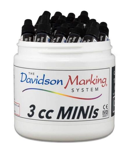
Mini Tissue Marking Dyes Black 3cc (pk/40)
Davidson Marking Systems® Mini Tissue Marking Dyes in Black colour. Tub of 40 x 3cc (3ml) bottles.
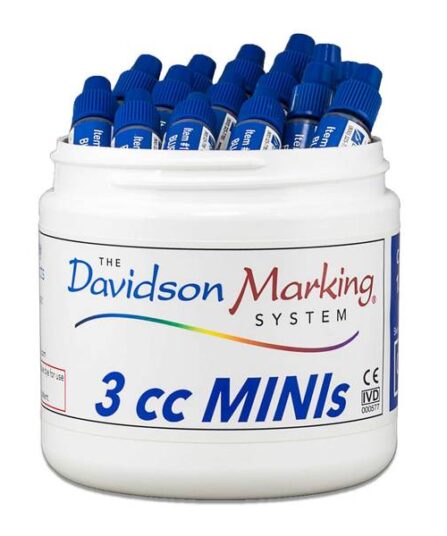
Mini Tissue Marking Dyes Blue 3cc (pk/40)
Davidson Marking Systems® Mini Tissue Marking Dyes in Blue colour. Tub of 40 x 3cc (3ml) bottles.
Tissue Marking Dye
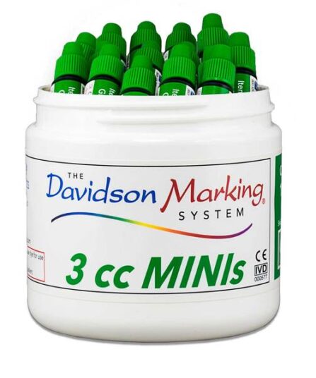
Mini Tissue Marking Dyes Green 3cc (pk/40)
Davidson Marking Systems® Mini Tissue Marking Dyes in Green colour. Tub of 40 x 3cc (3ml) bottles.
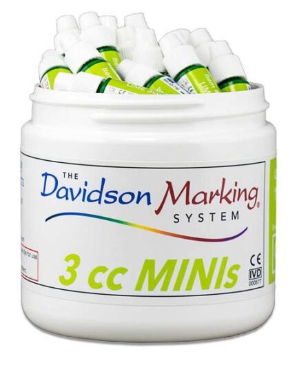
Mini Tissue Marking Dyes Lime Green 3cc (pk/40)
Davidson Marking Systems® Mini Tissue Marking Dyes in Lime Green colour. Tub of 40 x 3cc (3ml) bottles.
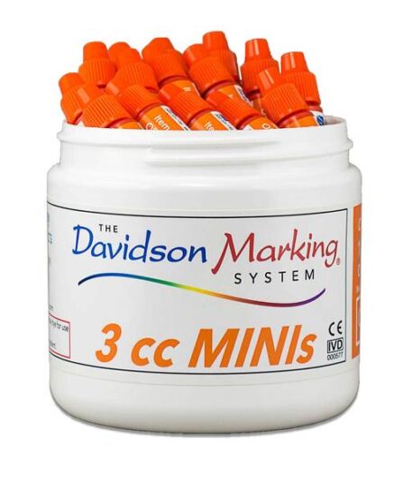
Mini Tissue Marking Dyes Orange 3cc (pk/40)
Davidson Marking Systems® Mini Tissue Marking Dyes in Orange colour. Tub of 40 x 3cc (3ml) bottles.
Histology
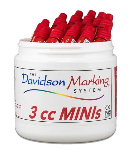
Mini Tissue Marking Dyes Red 3cc (pk/40)
Davidson Marking Systems® Mini Tissue Marking Dyes in Orange colour. Tub of 40 x 3cc (3ml) bottles.
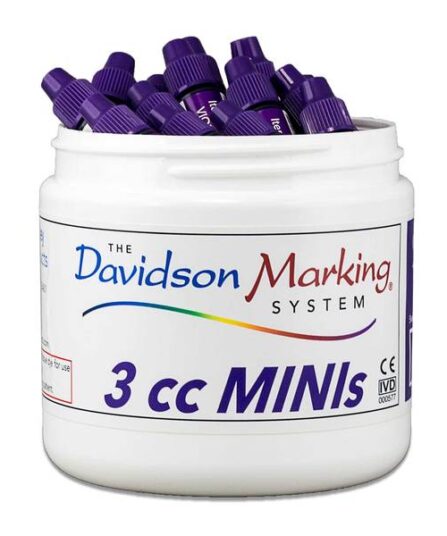
Mini Tissue Marking Dyes Violet 3cc (pk/40)
Davidson Marking Systems® Mini Tissue Marking Dyes in Violet colour. Tub of 40 x 3cc (3ml) bottles.
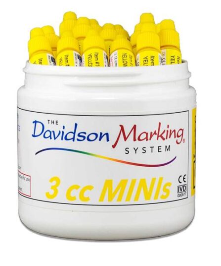
Mini Tissue Marking Dyes Yellow 3cc (pk/40)
Davidson Marking Systems® Mini Tissue Marking Dyes in Yellow colour. Tub of 40 x 3cc (3ml) bottles.
Stain Kits
Mucicarmine kit
Mucicarmine kit is often used to identify primary tumour sites, distinguishing mucin-negative undifferentiated squamous cell lesions from mucin-positive adenocarcinomas. It can also be used as indicative of diseases such as asthma, bronchitis, chronic obstructive pulmonary disease and cystic fibrosis.
Stain Kits
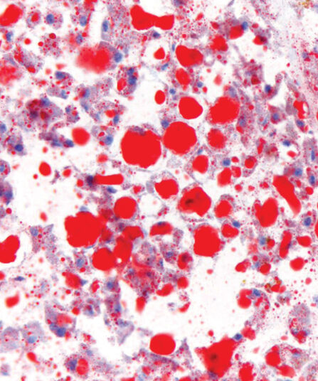
Oil Red O Kit
Four-reagent kit for selective staining and detection of fat cells and neutral fats according to Johnson. It can be used with frozen sections and fresh smears to detect obesity-linked pathologies such as dyslipidemia and diabetes type.
Stain Kits
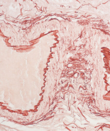
Orcein kit
Five-reagent kit for visualisation of hepatitis B surface antigen (HBsAg) seen as viral inclusion bodies in hepatocytes, for elastic fibres and copper associated protein in tissue sections. It can be used with frozen sections.
Stain Kits
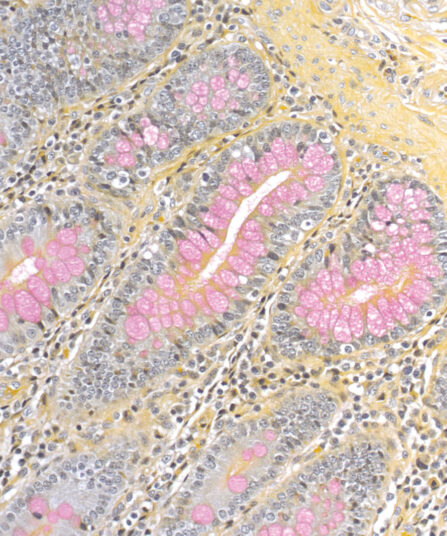
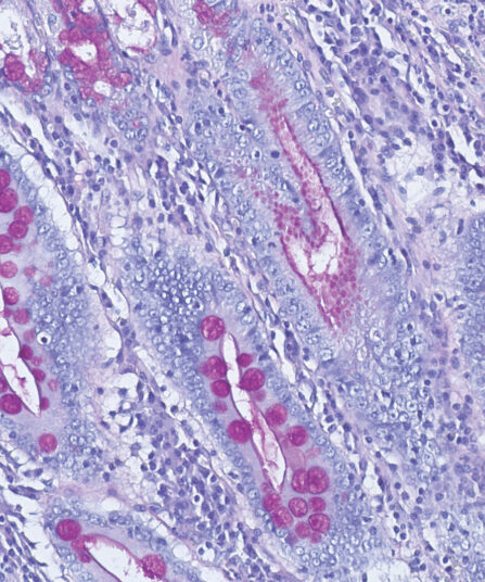
P.A.S. Diastase Kit
BioGnost’s P.A.S. Diastase kit is most commonly used for identifying glycogen in liver. Periodic acid enables the molecules containing glycol groups to create aldehydes affected by Schiff’s reagent staining them violet (magenta). Specific stains are created by applying the PAS method on unsubsti-tuted polysaccharides, mucoproteins and glycoproteins, glycolipids and phospholipids. Alpha-amylase enzyme (also known as diastasis) is used for differentiation between glycogen and other PAS-positive structures by dissolving 1→4 glycosidic bonds, causing the glycogen to remain unstained after the PAS reaction. BioGnost’s P.A.S. Diastase kit uses thermostable enzyme which does not require heating to +37°C to be active, but incubat-ing the section at +37°C is preferred in order to achieve better glycogen breakdown. The same tissue section is used as negative control for this reaction, but the sample is not treated using alpha-amylase.
For 100 tests.
Stain Kits
P.A.S. kit
Five-reagent Periodic Acid-Schiff kit for staining aldehydes, muccopolysaccharides, mucoproteins and lymphocytes according to Hotchkiss-McManus. P.A.S. staining may also be used for the demonstration of fungal organisms in tissue sections.
Stain Kits
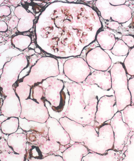
P.A.S.M. / Jones kit, stabilised
Seven-reagent Periodic Acid Silver Methenamine kit for staining kidney glomerular basement membranes. Kit includes red counterstain which provides visually rich contrast to target structures stained black.
Stain Kits
Paraldehyde Fuchsin kit
Seven-reagent kit according to Gomori for detecting pathological changes in elastic fibres. It also stains mast cell granules, beta granules in pancreatic islets, neurosecretory material, mast cell granules and beta cells in the pituitary gland.

Stains Reagents And Dyes