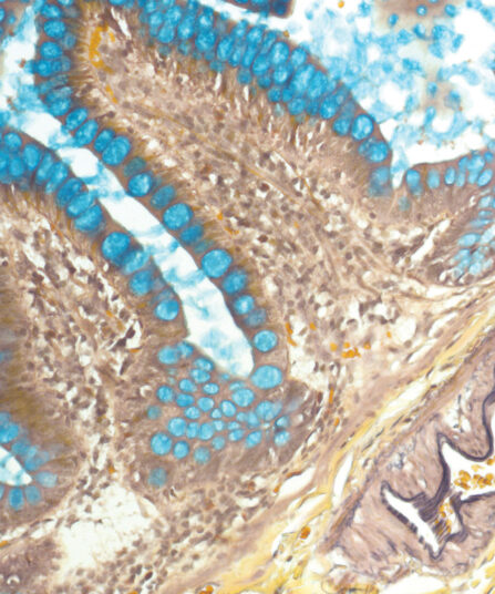
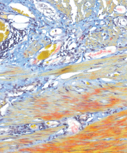
Masson-Goldner Trichrome kit is used for visualisation of muscles, collagen fibres, connective tissues, gametes, nuclei, neurofibrils, neuroglia, collagen, keratin intracellular fibrils and Golgi apparatus negative staining. Method of staining muscle and collagen fibres in tissues during which Fast Green F.C.F. dye binds to collagen making it turn distinct green. It is also used for visualisation of increased collagen build-up associated with functioning tissue being mistaken for scar tissue (liver sclerosis diagnosis), but also for differentiating smooth muscle fibres and collagens.
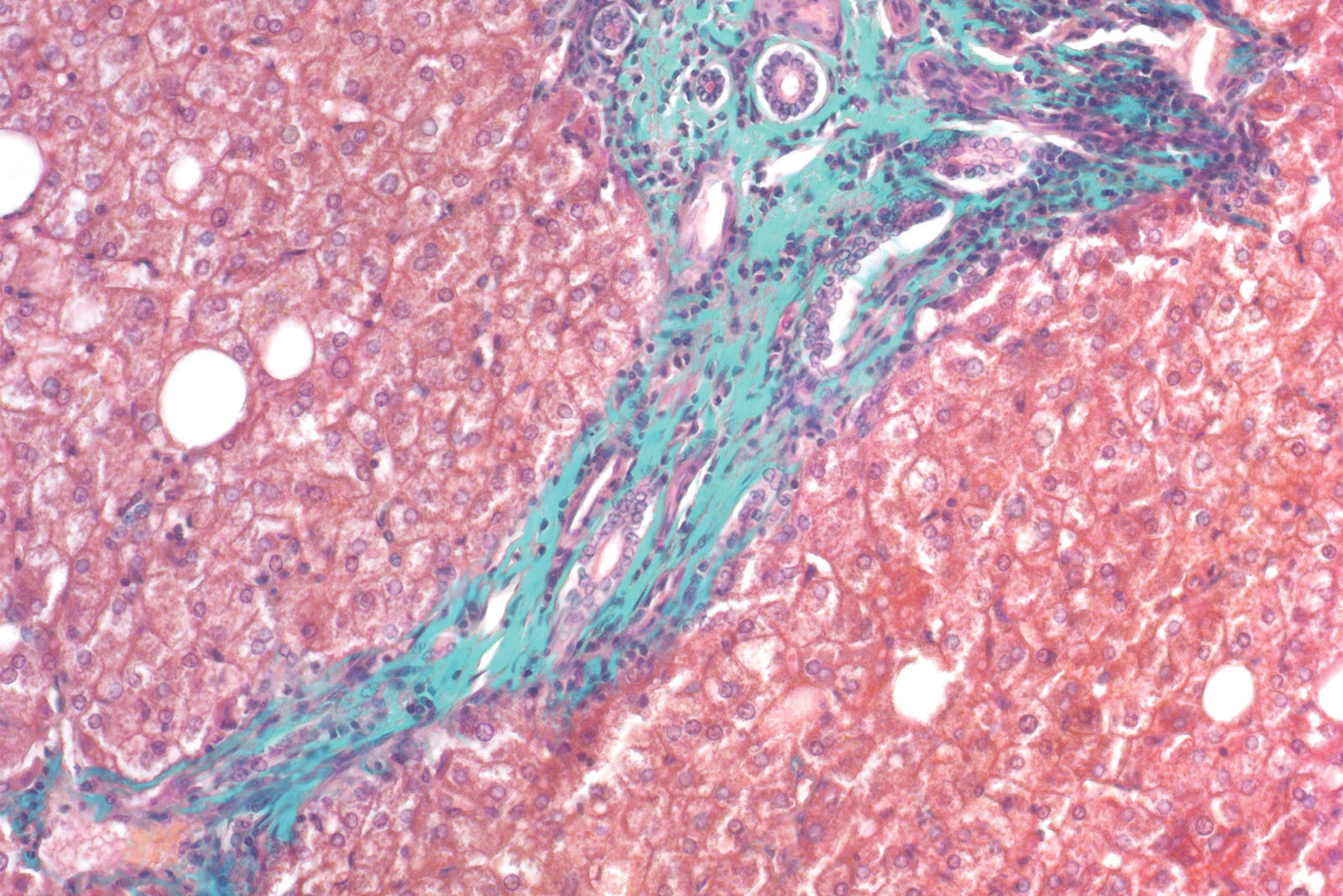
Masson-Goldner Trichrome kit
Seven-reagent kit for staining muscle and collagen fibres with green counterstain. It is also used for visualising gametes, nuclei, neurofibrils, glial cells, keratins, intercellular fibrils and for differentiation of smooth muscle fibres and collagens.
Description
Additional information
| Size | |
|---|---|
| Brand | |
| Stain pack | |
| Stain Category | Muscle and Connective Tissue |
Downloads
Related products
Stain Kits
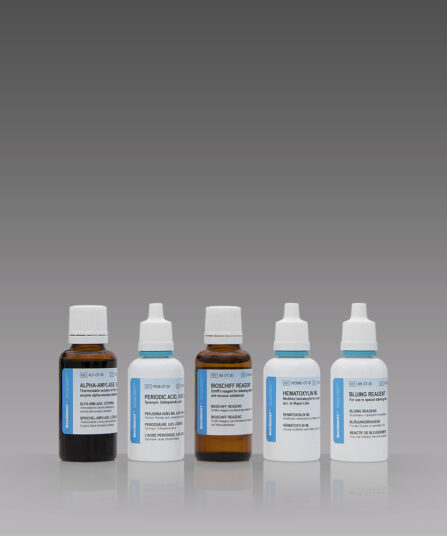
P.A.S. Diastase Kit
BioGnost’s P.A.S. Diastase kit is most commonly used for identifying glycogen in liver. Periodic acid enables the molecules containing glycol groups to create aldehydes affected by Schiff’s reagent staining them violet (magenta). Specific stains are created by applying the PAS method on unsubsti-tuted polysaccharides, mucoproteins and glycoproteins, glycolipids and phospholipids. Alpha-amylase enzyme (also known as diastasis) is used for differentiation between glycogen and other PAS-positive structures by dissolving 1→4 glycosidic bonds, causing the glycogen to remain unstained after the PAS reaction. BioGnost’s P.A.S. Diastase kit uses thermostable enzyme which does not require heating to +37°C to be active, but incubat-ing the section at +37°C is preferred in order to achieve better glycogen breakdown. The same tissue section is used as negative control for this reaction, but the sample is not treated using alpha-amylase.
For 100 tests.
Stain Kits
BioGram 4 kit
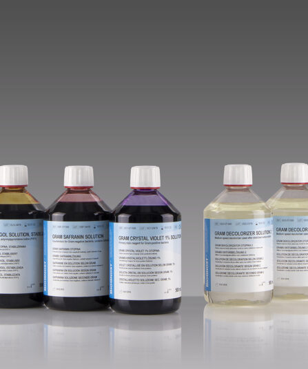
Four-reagent kit for identification of bacteria according to Gram. Kit contains Gram Crystal Violet 1% solution, stabilized Gram Lugol solution, double amount of Gram Decolorizer solution 2 and Gram Safranin solution as counterstain.
5×100 ml bottles

Stain Kits
TB-Stain Fluorescent Kit
Three-reagent kit for fluorescence-microscopic detection of acid-fast bacteria. Contains TB Auramine-Rhodamine reagent, double amount of TB Decolorizer Fluorescent and TB Permanganate reagent as counterstain.
4 x 100ml bottles.
Stain Kits
BioGram ECO kit
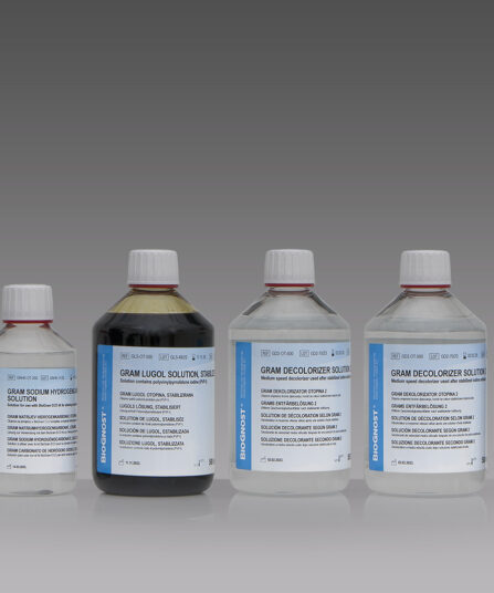
Four-reagent phenol-free kit for the identification of bacteria according to Gram. Kit contains Gram Crystal violet, phenol free reagent, Gram Sodium hydrogencarbon, solution, stabilized Gram Lugol solution, double amount of Gram Decolorizer solution 2 and Gram Safranin solution as counterstain.
2×50 mL+4×100 mL bottles

Stain Kits
TB-Stain Histo kit
Three-reagent kit for staining acid-fast bacteria (pathogenic mycobacteria) in histology sections, sputum, smears and culture smears according to Ziehl-Neelsen. Heating of the carbol-fuchsin solution is avoided in this protocol hence omitting the release of hazardous phenolic vapours.
Stain Kits
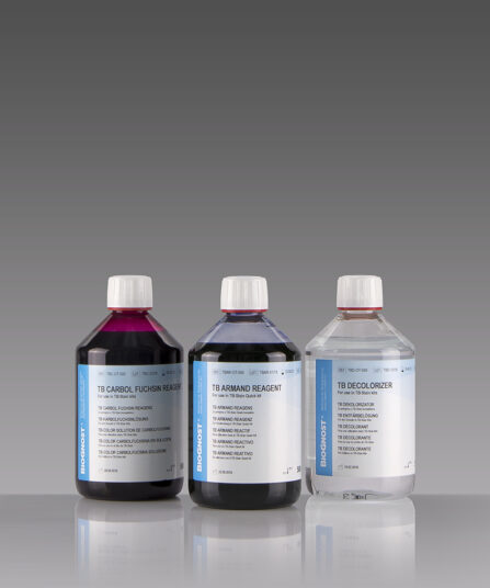
TB-Stain Quick Kit
Three-reagent kit for rapid staining of acid-fast bacteria using Kinyoun-Gabbett method. Contains TB Carbol Fuchsin reagent and TB Armand reagent as counterstain.
3 x 100ml bottles.
Stain Kits
Martius Scarlet Blue (MSB) kit
Seven-reagent kit used for fibrin visualisation, especially of older clusters. This method is a modification of Masson Trichrome and is ideal for studying connective tissue and vascular pathology.