
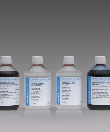
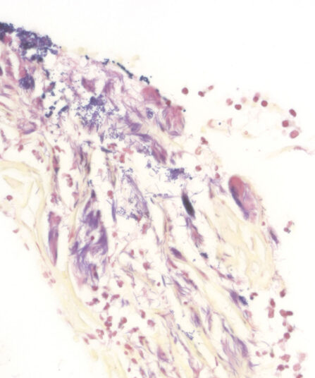
Many bacterial cells are easily stained by using simple dyes or Gram stain. However, a few bacterial strains, such as Mycobacteria and Nocardia cannot be stained using simple dyes (or, if successfully stained, the results may vary significantly). Cellular wall of the Mycobacteria strain contains a waxy substance – mycolic acid. Those are beta-hydroxy carboxylic acids with chains containing up to 90 carbon atoms. Its resistance to acidity is associated with mycolic acid chain length. In order to stain such strains, a higher concentration of dye or a longer period of heating is required. However, once stained, the dye is even more difficult to remove from the cells. Those bacteria are called acid-resistant because they maintain their primary colour even after decolourisation using acid alcohol (Carbol Fuchsin). Early laboratory diagnosis of tuberculosis is based on the interpretation of stained smears, and one of the best diagnostic methods is analysing sputum samples under a microscope. The most common and renowned method used for detecting the tuberculosis bacteria is staining according to Ziehl-Neelsen. This method uses Carbol Fuchsin as the main dye, acid alcohol as decolourisation medium and Methylene Blue solution as a contrasting dye. BioGnost’s TB-Stain Hot kit contains TB Carbol Fuchsin reagent, two packages of TB Decolouriser and Methylene Blue Loeffler reagent.
TB-Stain Histo kit
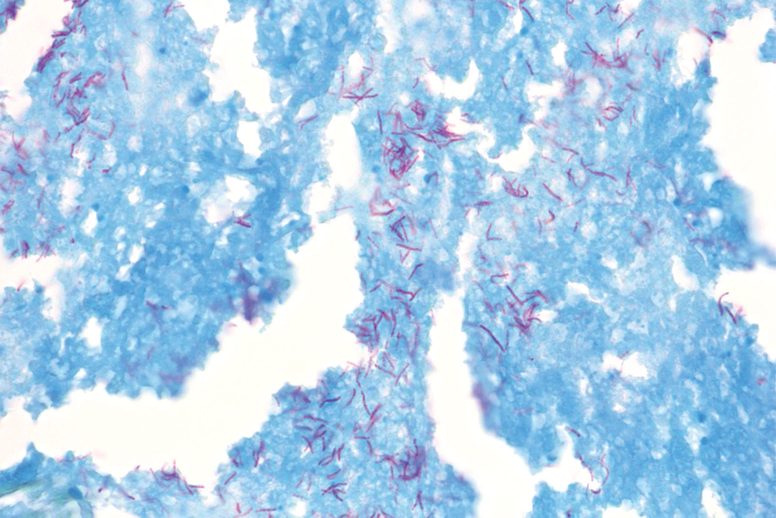
Three-reagent kit for staining acid-fast bacteria (pathogenic mycobacteria) in histology sections, sputum, smears and culture smears according to Ziehl-Neelsen. Heating of the carbol-fuchsin solution is avoided in this protocol hence omitting the release of hazardous phenolic vapours.
Description
Additional information
| Size | |
|---|---|
| Brand | |
| Stain pack | |
| Stain Category | Bacteria and Parasites, Fungi |
Downloads
Related products
Stain Kits
TB-Stain Quick Kit
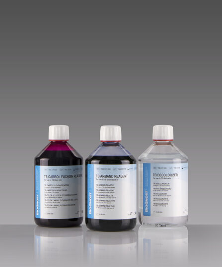
Three-reagent kit for rapid staining of acid-fast bacteria using Kinyoun-Gabbett method. Contains TB Carbol Fuchsin reagent and TB Armand reagent as counterstain.
3 x 100ml bottles.
Stain Kits
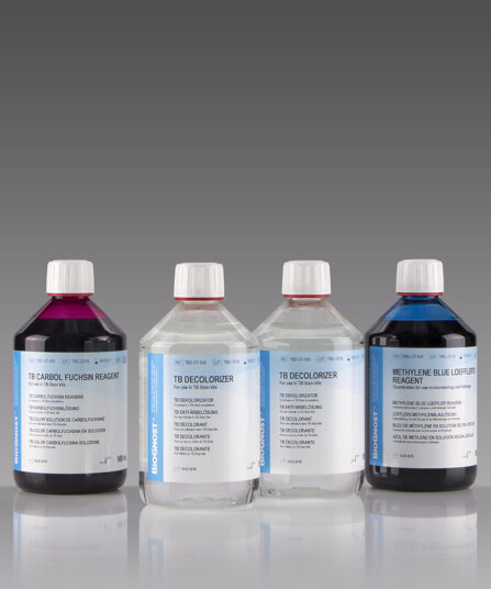
TB-Stain Hot Kit
Three-reagent kit for staining acid-fast bacteria. Contains TB Carbol Fuchsin reagent, double amount of TB Decolorizer and Methylene Blue Loeffler’s reagent as counterstain.
4 x 100ml bottles.
Stain Kits
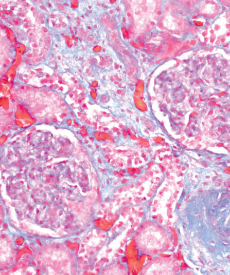
Gomori Trichrome kit
Five-reagent kit for staining muscle, collagen fibre and nuclei, contains blue counterstain. The kit can be used to contrast skeletal, cardiac or smooth muscle.
Stain Kits
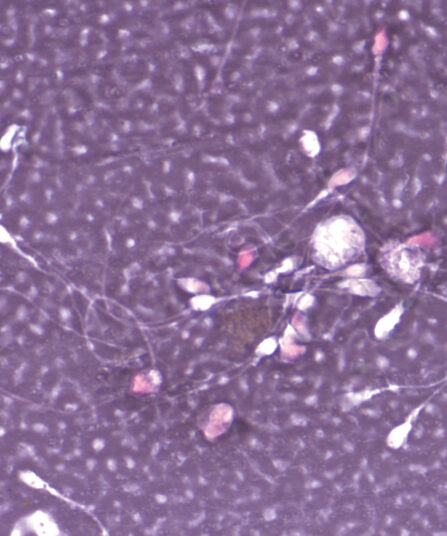
Eosin-Nigrosin Vital
Fast detection (one-step detection) of sperm vitality and visualisation of dead and living sperm cells with one reagent. A simple, easy and fast method for semen analysis.
Stain Kits
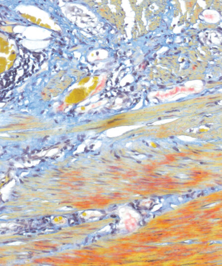
Martius Scarlet Blue (MSB) kit
Seven-reagent kit used for fibrin visualisation, especially of older clusters. This method is a modification of Masson Trichrome and is ideal for studying connective tissue and vascular pathology.
Stain Kits
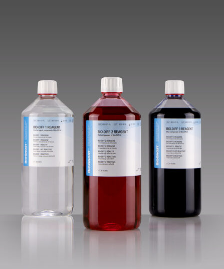
Bio-Diff Kit 3 X 1L
Three-reagent kit that contains fixative agent, red and blue components for fast and effective staining. Each kit contains buffer tablets for consistent staining results.
3×100 ml bottles
Stain Kits
TB-Stain ECO Kit
Three-reagent phenol-free kit for staining acid-fast bacteria. Contains TB-Fuchsin reagent, double amount of TB Decolorizer and Methylene Blue Loeffler’s reagent as counterstain.
5 x 100ml bottles.
Stain Kits
BioGram Histo kit
Five-reagent kit for identification of bacteria according to Gram. For differentiation between Gram-positive and Gram-negative bacteria in histology sections.