Histology, the study of microscopic tissues, involves several steps before the sample is ready to go under the microscope.
Histology lab managers place very high importance on the sample preparation process, ensuring the sections produced contain all the microscopic information required to obtain the correct diagnosis.
Here are some of the steps involved in the preparation of a histology sample which can differ between labs and constantly evolves as new processes, techniques, and equipment are developed to improve results.
Step 1: Fixation
Fixation is where the tissue sample is preserved and stabilised to prevent decay and maintain cellular structures. Fixatives such as formalin are used to achieve this. The sample is immersed in a liquid fixing agent (fixative) the most used being a 10% buffered formalin solution.
Specimens remain in the fixative solution long enough for the fixative to penetrate every part of the tissue, plus an additional period to allow the chemical reactions of fixation to reach equilibrium, generally around 6 and 24 hours.
Step 2: Dehydration
After fixation, the sample needs to be dehydrated to remove water and replace it with a solvent that is miscible with the embedding medium (paraffin wax).
The tissue is passed through a series of increasing concentrations of alcohol (e.g., 70%, 80%, 95%, and 100%) to gradually remove water from the tissue.
An example dehydration sequence for specimens no more than 4mm thick would be:
- 70% ethanol 15 min
- 90% ethanol 15 min
- 100% ethanol 15 min
- 100% ethanol 15 min
- 100% ethanol 30 min
- 100% ethanol 45 min
Following this, all but a tiny residue of water will have been removed from the specimen.
Step 3: Clearing
Following dehydration, the tissue is immersed in a clearing agent (such as xylene) to remove the alcohol and render the tissue transparent.
Clearing helps to improve the penetration of the embedding medium.
Step 4: Infiltration
Infiltration involves impregnating the dehydrated and cleared tissue with an embedding medium, typically paraffin wax.
The tissue is placed in melted paraffin wax, and through a process called impregnation, the paraffin replaces the clearing agent (xylene or a xylene alternative), ensuring the tissue is embedded uniformly.
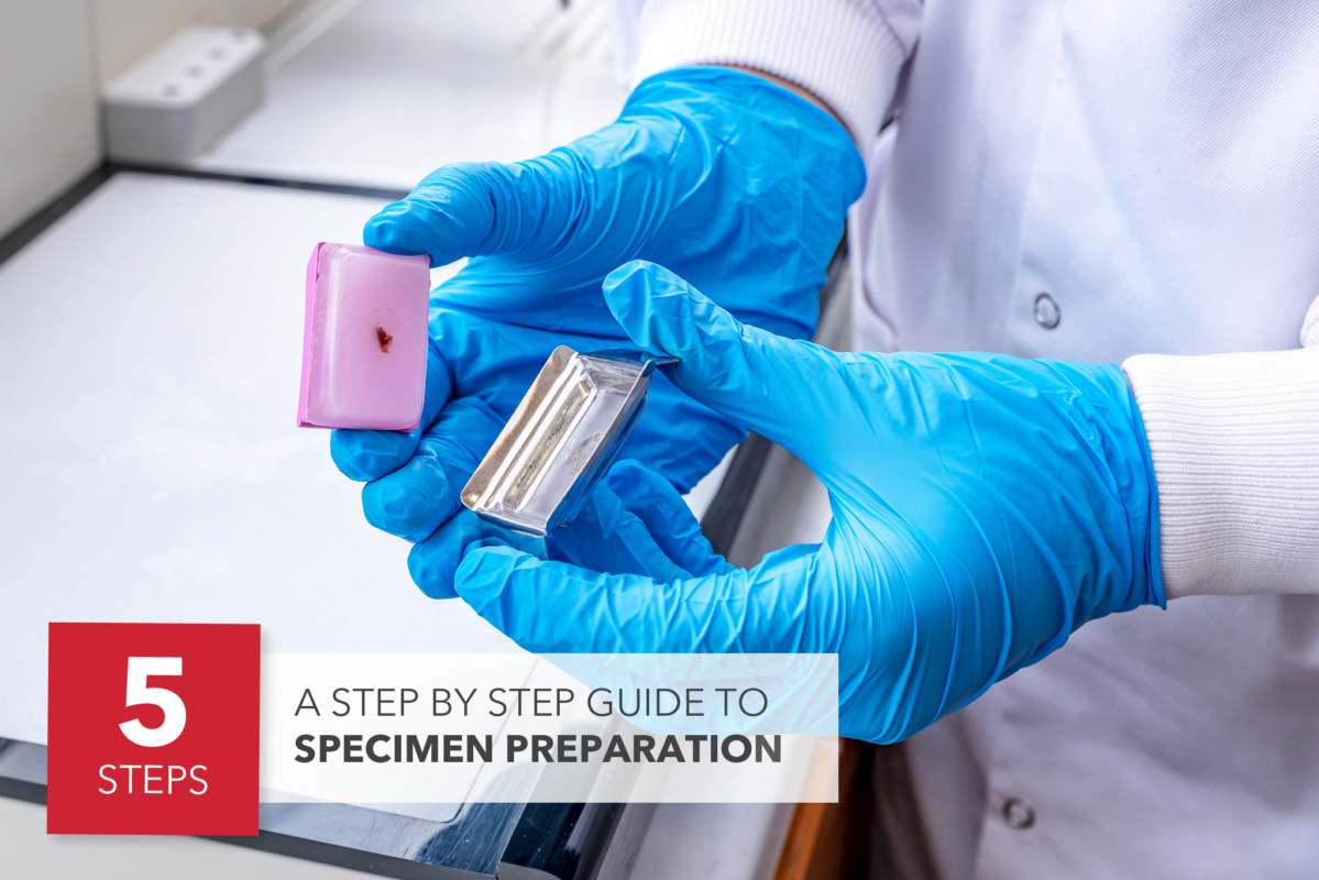
Step 5: Embedding
The infiltrated tissue is now ready to be formed into a “block” which is transferred to an embedding centre.
The infiltrated tissue sample is placed into a tissue mold (mold size dependent on the sample size) containing fresh molten paraffin wax. The tissue is oriented appropriately, ensuring the sectioning plane is ideal for subsequent microscopic examination. The paraffin is allowed to solidify, forming a “block” that contains the tissue.
To embed the infiltrated tissue, it is important to plan the orientation: Based on the features of interest and the tissue type, determine the ideal orientation for sectioning, taking into consideration the anatomical structure, the desired orientation of the sections (longitudinal, transverse, etc.), and the orientation required for specific staining techniques.
Place the tissue sample in a histology tissue mold, ensuring that it is positioned appropriately based on the desired orientation. The mold should be large enough to accommodate the tissue and provide support during processing.
Fill the mold with molten paraffin wax ensuring that the tissue is completely submerged in the wax and properly orientated using forceps or heated forceps for easier orientation.
Let the wax solidify around the tissue sample. This can be achieved by cooling the mold on a cold plate.
A cassette is placed on top, and an embedding bead is placed in the corner of the cassette before the mold is topped up with molten paraffin wax and again placed on the cold plate or put in the fridge to solidify.
Choosing the right embedding medium (paraffin wax) is crucial in this process to obtain the best sections.
The embedded tissue block is now ready to cut into thin sections using a precision microtome.