
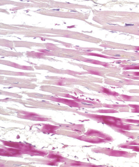
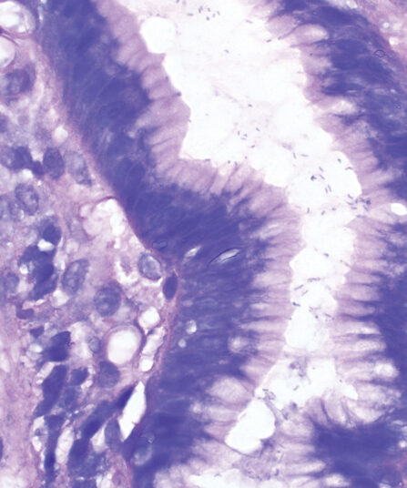
Van Gieson trichrome kit is used for staining collagen, muscle tissue, keratinized epithelium, cytoplasm, glial fibres, and erythrocytes. Fuchsin Acid Van Gieson is a component of the kit and it contains two dyes (acid fuchsin, picric acid) that simultaneously stain different tissue structures. Acid fuchsin stains collagen fibres intensive red while picric acid stains muscle fibres, erythrocytes and glial fibres yellow. Amyloids, hyalin, colloid and mucosa are stained in nuances between red and yellow. Hematoxylin, Weigert A and Ferri reagent make up Weigert hematoxylin that creates stable cellular nuclei colouration.
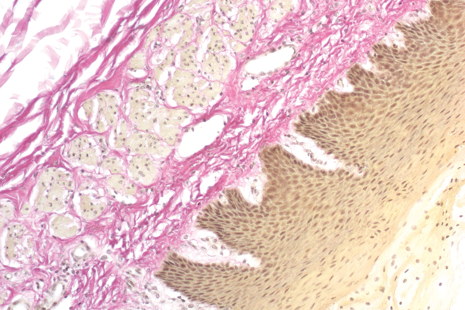
Van Gieson Trichrome kit
Three-reagent kit for staining collagen fibres, muscle tissue, keratinized epithelium, cytoplasm, glial fibres and erythrocytes. Used for differentiation between collagen and smooth fibres in tumours and various other diseases.
Description
Additional information
| Size | |
|---|---|
| Brand | |
| Stain pack | |
| Stain Category | Muscle and Connective Tissue |
Downloads
Related products
Stain Kits
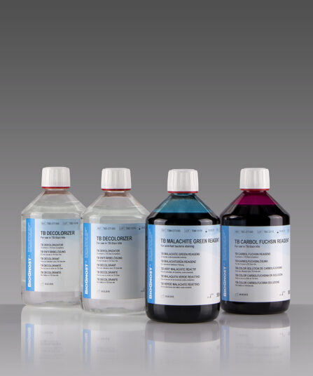
TB-Stain Cold Kit
Three-reagent kit for staining acid-fast bacteria according to Kinyoun. Contains TB Carbol Fuchsin reagent, double amount of TB Decolorizer and TB Malachite Green reagent as counterstain.
4 x 100ml bottles.
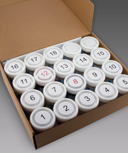
HE Rapid Staining kit- frozen and paraffin sections
Ready-to-use eight-reagent kit (in 16 containers that can be used as staining jars) for rapid HE staining of frozen and paraffin tissue sections in histopathology. Contains xylene substitute as clearing agent and xylene substitute-based medium for permanent section covering.
For 100 tests.
Stain Kits
H.B.F.P. kit
Three-reagent Hematoxylin-Basic Fuchsin-Picric acid staining kit for detection of cardiac muscle changes after ischemia or myocardial infarction. H.B.F.P. kit is a non-enzymatic histochemical technique for detection of early myocardial ischemia with vivid contrast.
Stain Kits
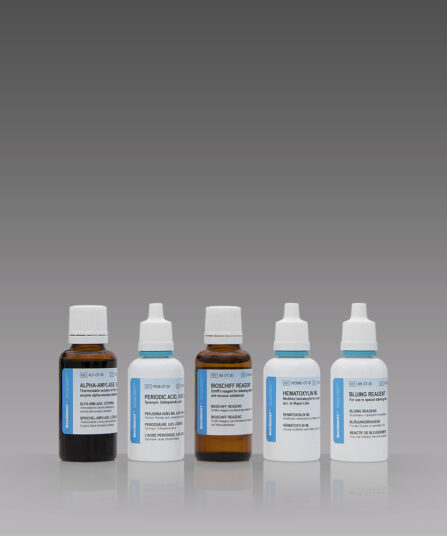
P.A.S. Diastase Kit
BioGnost’s P.A.S. Diastase kit is most commonly used for identifying glycogen in liver. Periodic acid enables the molecules containing glycol groups to create aldehydes affected by Schiff’s reagent staining them violet (magenta). Specific stains are created by applying the PAS method on unsubsti-tuted polysaccharides, mucoproteins and glycoproteins, glycolipids and phospholipids. Alpha-amylase enzyme (also known as diastasis) is used for differentiation between glycogen and other PAS-positive structures by dissolving 1→4 glycosidic bonds, causing the glycogen to remain unstained after the PAS reaction. BioGnost’s P.A.S. Diastase kit uses thermostable enzyme which does not require heating to +37°C to be active, but incubat-ing the section at +37°C is preferred in order to achieve better glycogen breakdown. The same tissue section is used as negative control for this reaction, but the sample is not treated using alpha-amylase.
For 100 tests.
Stain Kits
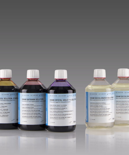
BioGram 4 kit
Four-reagent kit for identification of bacteria according to Gram. Kit contains Gram Crystal Violet 1% solution, stabilized Gram Lugol solution, double amount of Gram Decolorizer solution 2 and Gram Safranin solution as counterstain.
5×100 ml bottles
Stain Kits
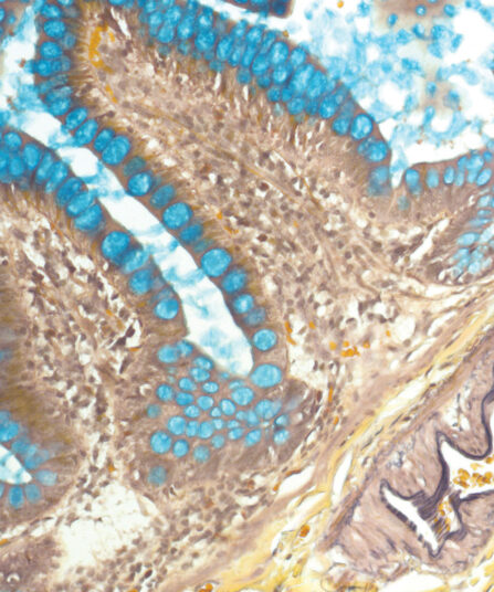
Giemsa HP kit
Four-reagent kit for staining Helicobacter pylori in gastroscopic sections according to Lennart. Advantages of this method for detecting H. pylori are sensitive and reproducible results and easy performance.
Stain Kits
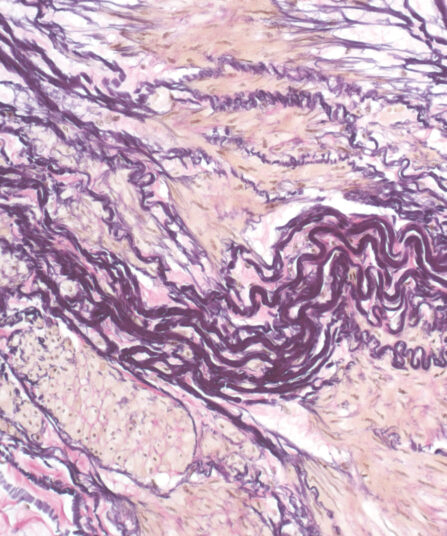
Elastica-Van Gieson kit
Four-reagent kit for staining elastic fibres and differentiation between elastic tissue, collagen and other types of connective tissue. The rapid method enables a satisfactory result with shorter section staining time.