Many bacterial cells are easily stained by using simple dyes or Gram stain. However, a few strains of bacteria, such as Mycobacteria and Nocardia cannot be stained using simple dyes (the results may vary significantly if successfully stained). Cellular wall of the Mycobacteria strain contains waxy substance – mycolic acid. Those are beta-hydroxy carboxylic acids with chains containing up to 90 carbon atoms. Its resistance to acidity is associated with mycolic acid chain length. In order to stain such strains, a higher concentration of dye or a longer period of heating is required. However, once stained, the dye is even more difficult to remove from the cells. Those bacteria are called acid-resistant because they maintain their primary color even after decoluorisation using acid alcohol (Carbol Fuchsin). Early laboratory diagnosis of tuberculosis is based on the interpretation of stained smears, and one of the best diagnostic methods is analyzing sputum sample under a microscope. The method according to Kinyoun is an alternative to the Ziehl-Neelson method of detecting tuberculosis bacteria. The Kinyoun method does not require heating the glass slide containing the sample. This method uses Carbol Fuchsin as the main dye, acid alcohol as decolourisation medium and Malachite Green solution as a contrasting dye. BioGnost’s TB-Stain Cold kit contains TB Carbol Fuchsin reagent and two packages of TB Decolorizer and Malachite Green reagent.
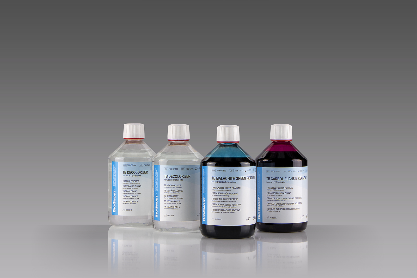
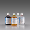
TB-Stain Cold Kit
Three-reagent kit for staining acid-fast bacteria according to Kinyoun. Contains TB Carbol Fuchsin reagent, double amount of TB Decolorizer and TB Malachite Green reagent as counterstain.
4 x 100ml bottles.
Related products

Stain Kits
Bio-Diff kit 3 x 500ml
Three-reagent kit that contains fi xative agent, red and blue components for fast and effective staining. Each kit contains buffer tablets for consistent staining results.
Stain Kits
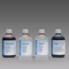
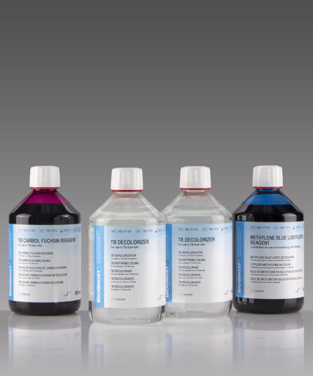
TB-Stain Hot Kit
Three-reagent kit for staining acid-fast bacteria. Contains TB Carbol Fuchsin reagent, double amount of TB Decolorizer and Methylene Blue Loeffler’s reagent as counterstain.
4 x 100ml bottles.
Stain Kits
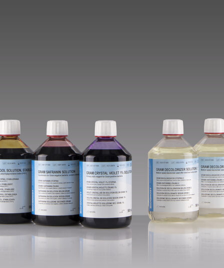
BioGram 4 kit
Four-reagent kit for identification of bacteria according to Gram. Kit contains Gram Crystal Violet 1% solution, stabilized Gram Lugol solution, double amount of Gram Decolorizer solution 2 and Gram Safranin solution as counterstain.
5×100 ml bottles
Stain Kits
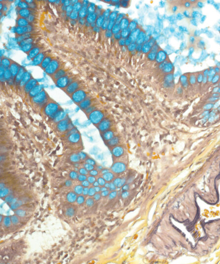
Giemsa HP kit
Four-reagent kit for staining Helicobacter pylori in gastroscopic sections according to Lennart. Advantages of this method for detecting H. pylori are sensitive and reproducible results and easy performance.
Stain Kits
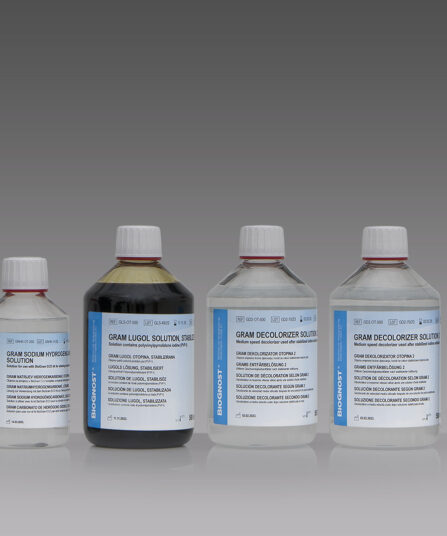
BioGram ECO kit
Four-reagent phenol-free kit for the identification of bacteria according to Gram. Kit contains Gram Crystal violet, phenol free reagent, Gram Sodium hydrogencarbon, solution, stabilized Gram Lugol solution, double amount of Gram Decolorizer solution 2 and Gram Safranin solution as counterstain.
2×50 mL+4×100 mL bottles
Stain Kits
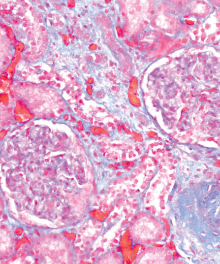
Gomori Trichrome kit
Five-reagent kit for staining muscle, collagen fibre and nuclei, contains blue counterstain. The kit can be used to contrast skeletal, cardiac or smooth muscle.
Stain Kits
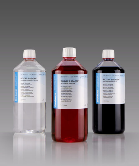
Bio-Diff Kit 3 X 1L
Three-reagent kit that contains fixative agent, red and blue components for fast and effective staining. Each kit contains buffer tablets for consistent staining results.
3×100 ml bottles