MICROTOMY
Microtomy is the process followed to prepare thin slices of biological tissues for microscopic examination in histology. Thin, uniformed sections are necessary to obtain, enabling pathologists to examine and study the microscopic structure of tissues and cells under a microscope.
More...
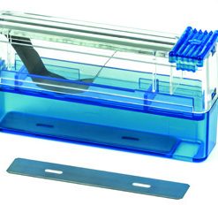
To achieve the thin sections required histopathologists use a rotary microtome designed to cut embedded specimen blocks. The microtome trims the front layer of the paraffin-embedded block with a sharp blade as the block manually or automatically moves up and down against the blade. This leaves behind a thin specimen section/ribbon ranging from 3μm to 100 μm that can then be mounted on a microscope slide.
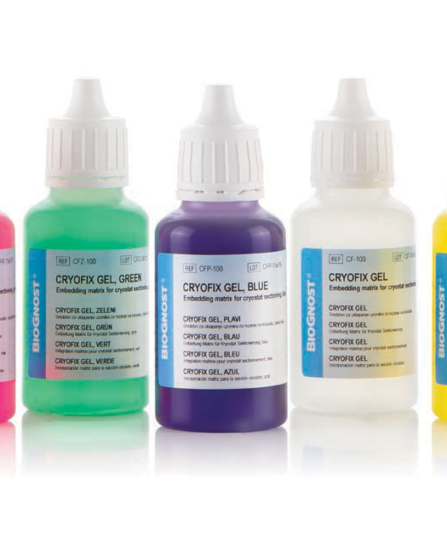
Paraffin-embedded tissue is sectioned using a manual, semi-automatic or fully automatic rotary microtome, whereas fresh tissue can be immediately sectioned using a Cryostat where the specimen is mounted onto a metal chuck and placed within the cryochamber to freeze.
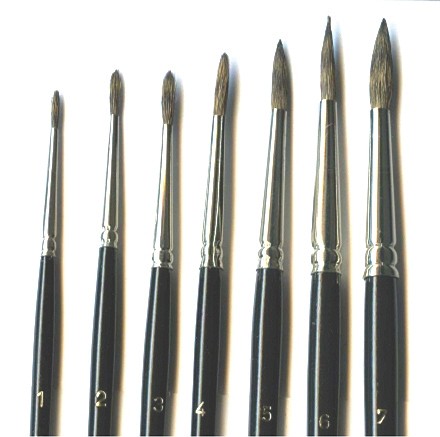
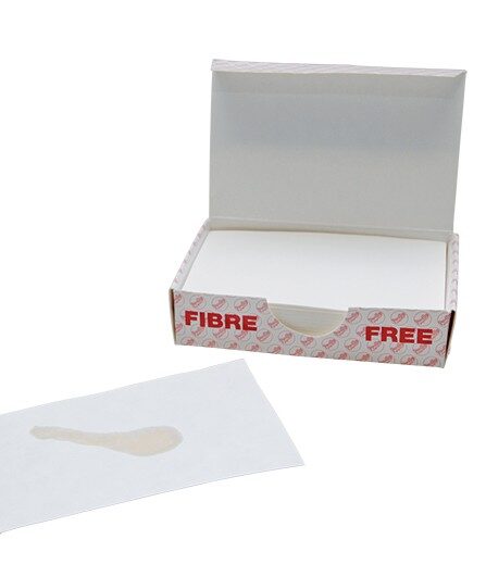
The microtome/cryostat will produce a ribbon on sections that are transferred into a water bath, removing any wrinkles and flattening the section, ready to be shortened with forceps, mounted onto microscope slide, ready for staining.
show less