
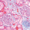
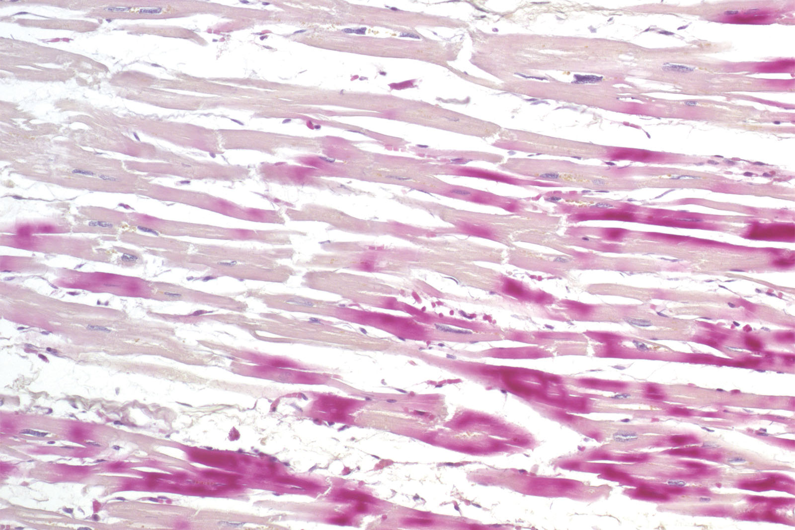
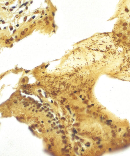
Histological diagnosis of ischemia in the early phase of myocardial infarction using the standard hematoxylin-eosin histological methods and a light microscope is exceptionally delicate. The reason for that is minimal histopathological changes occurring on the cardiac muscle during the first six hours of symptoms. However, staining the section using the kit consisting of hematoxylin, basic fuchsin and picric acid enables a histological overview of early changes on the cardiac muscle caused by ischemia or myocardial infarction.
H.B.F.P. kit
Three-reagent Hematoxylin-Basic Fuchsin-Picric acid staining kit for detection of cardiac muscle changes after ischemia or myocardial infarction. H.B.F.P. kit is a non-enzymatic histochemical technique for detection of early myocardial ischemia with vivid contrast.
Description
Additional information
| Size | |
|---|---|
| Brand | |
| Stain pack | |
| Stain Category | Muscle and Connective Tissue |
Downloads
Related products

Stain Kits
TB-Stain Histo kit
Three-reagent kit for staining acid-fast bacteria (pathogenic mycobacteria) in histology sections, sputum, smears and culture smears according to Ziehl-Neelsen. Heating of the carbol-fuchsin solution is avoided in this protocol hence omitting the release of hazardous phenolic vapours.
Stain Kits
Martius Scarlet Blue (MSB) kit
Seven-reagent kit used for fibrin visualisation, especially of older clusters. This method is a modification of Masson Trichrome and is ideal for studying connective tissue and vascular pathology.
Stain Kits
Mallory Trichrome kit
Three-reagent staining kit for connective tissue visualisation and detection of collagen, cartilage, muscle, elastic fibres, mucous, pituarity cells, reticulum, bones, amyloid and erythrocytes.
Martius Scarlet Blue (MSB) kit, 6x100ml+1x250ml
Seven-reagent kit used for fibrin visualisation, especially of older clusters. This method is a modification of Masson Trichrome and is ideal for studying connective tissue and vascular pathology.

Stain Kits
TB-Stain Fluorescent Kit
Three-reagent kit for fluorescence-microscopic detection of acid-fast bacteria. Contains TB Auramine-Rhodamine reagent, double amount of TB Decolorizer Fluorescent and TB Permanganate reagent as counterstain.
4 x 100ml bottles.
Stain Kits
Warthin Starry kit
Five-reagent kit for staining Spirochaeta, Helicobacter pylori, Microsporidia and Legionella pneumophila. The kit contains 12 jars with gelatine that enables both incubation and staining of sections, as well as other reagents that enable precipitation of silver on the bacterial surface. The bacteria are found in the mucus of the surface epithelium, in the apical gastric glands and in the gastric mucosa.

Stains Reagents And Dyes
Stain Kits
P.A.S. Diastase Kit
BioGnost’s P.A.S. Diastase kit is most commonly used for identifying glycogen in liver. Periodic acid enables the molecules containing glycol groups to create aldehydes affected by Schiff’s reagent staining them violet (magenta). Specific stains are created by applying the PAS method on unsubsti-tuted polysaccharides, mucoproteins and glycoproteins, glycolipids and phospholipids. Alpha-amylase enzyme (also known as diastasis) is used for differentiation between glycogen and other PAS-positive structures by dissolving 1→4 glycosidic bonds, causing the glycogen to remain unstained after the PAS reaction. BioGnost’s P.A.S. Diastase kit uses thermostable enzyme which does not require heating to +37°C to be active, but incubat-ing the section at +37°C is preferred in order to achieve better glycogen breakdown. The same tissue section is used as negative control for this reaction, but the sample is not treated using alpha-amylase.
For 100 tests.