MICROSCOPY
Microscopy is the term used for the process of using a microscope to observe objects and details that are too small to be seen with the naked eye. Microscope lenses enable you to magnify and resolve the structures or features of a sample or object, making them visible and understandable.
More...
Microscopy plays a pivotal part in patient diagnosis across all pathological specialities, enabling pathologists to identify abnormalities, fine structures, colour and the number of cells.
There’s a wide variety of different microscopy techniques practised across the labs that are required for different purposes and produce very different images of a sample.
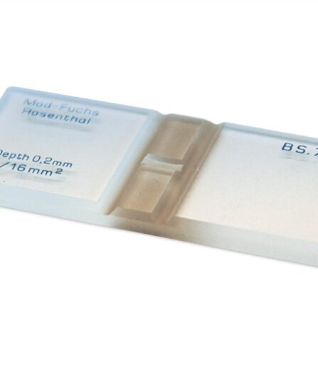
Optical or Light Microscopy: is where light from a source is transmitted and passed through the sample that is on a microscope slide to enlarge and magnify the image of the sample. Optical microscopy can be performed on living samples, making it possible to see cells migrate and divide in live action.
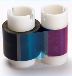
Fluorescence Microscopy: is a type of light microscopy used to image samples that fluoresce. Light of one wavelength is used to excite the fluorescent molecules, and the light of a different wavelength that they emit is then collected and used to form a picture.
Many fluorescent dyes can be used to stain structures such as fluorescein or rhodamine. One powerful method is the combination of antibodies coupled to a fluorophore as in immunostaining.
Of all the microscopy techniques, fluorescence is perhaps the most versatile. The fluorochromes designed for fluorescence microscopy permit the labelling of living cells, tissues, fixed and frozen sections. Fluorescence microscopy displays acid-fast bacilli labelled with auramine, detects autoantibodies, and enables the labelling of specific markers, cytoskeleton and nuclei.
Electron Microscopy: can be used to examine not only whole cells, but also the subcellular structures and compartments within them. Samples must be placed under vacuum and require an extensive fixation process. Therefore, live cells cannot be imaged this way.
There are two types of electron microscopy:
Transmission Electron Microscope (TEM): Allows for detailed imaging of internal structures of cells and tissues. This is most commonly used in diagnostic pathology.
Scanning Electron Microscope (SEM): Provides 3D surface images of specimens.
show less