H&E staining, also known as Hematoxylin and Eosin staining, is a commonly used staining technique in histology and pathology. The stains and protocol used provide a detailed picture of the microanatomy of organs and tissues. When it is properly performed it has the ability to demonstrate a wide range of normal and abnormal cell and tissue components and yet it is a relatively simple stain to carry out on paraffin or frozen sections. In histopathology, a high proportion of diseases can be diagnosed by using an H&E stain alone.
Hematoxylin
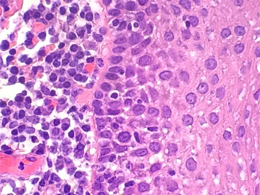
Hematoxylin precisely stains nuclear components with an affinity for acidic components. This includes staining heterochromatin, cell nuclei and other basophilic structures, such as the nucleoli and rough endoplasmic reticulum, in shades of blue-purple.
The type of mordant influences the final colour. The most common mordant used in routine histology is Aluminium Ammonium Sulphate (alum). This mordant causes the nuclei to be red in colour, which is then changed a blue colour when the sample is later rinsed. However, the depth of the colouration is related to the amount of DNA in the Nuclei and the length of time the samples spend in the stain.
Eosin
Esion, on the other hand, is an acidic dye that stains structures with an affinity for basic components. It stains cytoplasm and extracellular structures, such as collagen, elastic fibres, muscle fibres and red blood cells in shades of pink. Eosin is the most commonly used counterstain that distinguishes between the cytoplasm and nuclei of cells. Eosin displays different shades of pink for different types of connective tissue fibres.
Types of H&E Staining
There are typically three types of H&E staining: progressive, modified progressive, and regressive.
Progressive staining is when Hematoxylin is added to the tissue without being followed by a differentiator to remove excess dye. Because there is no differentiation step, background staining can occur, especially with charged or treated slides. This means noncellular material, such as mucin, becomes stained with the Hematoxylin which can be an indicator of well-differentiated tumours.
In contrast, regressive and modified progressive staining uses a differentiator. The strength of the differentiator will impact the staining. If the differentiator is too strong, it will remove more Hematoxylin and will make the nuclei pale. Too much time in a properly prepared differentiator will also remove more Hematoxylin and will ultimately under-stain the nuclei.
3 Benefits of H&E Staining
- Hematoxylin and Eosin staining is used as a type of control for all immunohistochemical staining to show the tissue was correctly processed and enables the histopathologist to make a disease diagnosis.
- H&E serves as a popular background stain in immunohistochemistry (IHC). When using an antibody to detect a specific protein through IHC, a background stain such as H&E is used to simultaneously visualise the cells where the protein is being detected.
- Before ‘special stains’ are applied, a tissue specimen is initially examined with H&E to determine the requirement for special stains in order to provide further analysis of the sample.
Steps of H&E Staining
Once your tissue samples have gone through the processes of Fixation, Dehydration, Clearing Infiltration/Embedding and Sectioning the sample is then ready to be stained using the following steps:
- Deparaffinization: Sections on slides are deparaffinized by immersing them in Xylene or another clearing agent to remove the embedding medium.
- Rehydration: The tissue samples are rehydrated by passing them through decreasing concentrations of alcohol to restore water content.
- Hematoxylin Staining: Sections are stained with Hematoxylin, staining the nuclei and some other elements a reddish-purple colour. The sample is then rinsed under tap water and then “blued” by treatment with a weakly alkaline solution. This step converts the Hematoxylin to a dark blue colour.
- Differentiation: Sections are treated with an acid alcohol or a weak acid solution to differentiate the stain, removing excess Hematoxylin and non-specific background staining from the cytoplasm and other cellular components.
- Eosin Staining: Sections are stained with Eosin that binds to basic components in the cytoplasm, nonnuclear elements, providing a pink to red colour.
Tip
Overstaining with Hematoxylin can give the illusion of under-stained Eosin, just as overstaining with Eosin can cause the Hematoxylin to appear lighter than it actually is. So, when optimising the stain, make sure to only edit the time of one of the components. This technique will help eliminate the need to spend additional time adjusting the stain.
- Dehydration and Clearing: Sections are dehydrated through increasing concentrations of alcohol and cleared with Xylene.
- Mounting: Sections are mounted with a mounting medium and covered with a coverslip for microscopic examination.