Tissue Marking Dyes are permanent inks used to mark orientations or surfaces of tissue specimens submitted for histopathology. This allows histopathologists to maintain proper orientation when examining the specimen under the microscope.
How do they work?
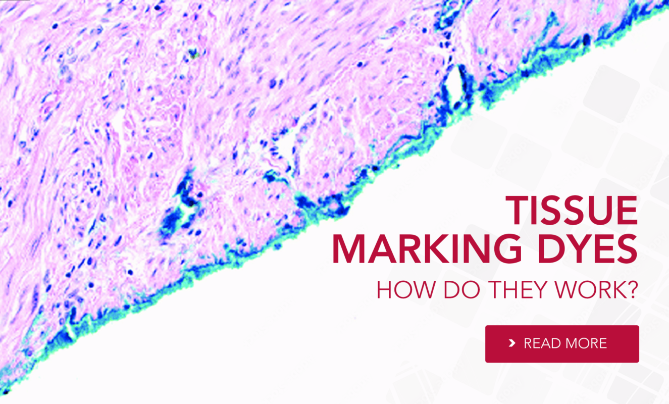
Tissue marking dyes have cationic (positively) charged properties, which are attracted to the negatively charged, anionic tissue components forming strong ionic bonds.
Once attached, these bonds are permanent and remain resistant to the tissue processing chemicals and other factors, maintaining their pigmentation. Some tissue components are amphoteric (neutrally charged) and will simply take up the dye by adsorption.
Once the tissue is marked with the dye, you can identify the boundaries of the tumour and assess the margins in the specimen. The correct orientation allows you to know if the margins identified are superior or inferior. Pathologists can use the marked margins as a guide to precisely evaluate the extent of the tumour and determine whether the margins are clear of cancer cells.
How Pathologists use Tissue Marking Dyes during Mohs?v
Mohs micrographic surgery is the surgical technique used for the removal of skin cancers, especially those with high recurrence rates or located in cosmetically sensitive areas.
Tissue Marking Dyes are commonly used during Mohs Surgery, once the initial tumour has been removed, the dyes are applied to the outer edges of the extracted tissue (peripheral and deep margins) to mark the surgical margins, to indicate the orientation and the specific locations on the excised tissue.
The marked tissue is processed and sectioned in a cryostat perpendicular to the marked margins. When the section is mounted onto the slide the dyes help maintain the orientation with the original surgical site. The pathologists examine the sections layer by layer and the dyes enable precise mapping and identification of the tissue layers. Despite the applied histological stains, the marked margins guide pathologists in identifying cancerous cells at the edges of the tissue sections.
The information provided by the marked margins helps to assess the success of the surgery and the completeness of the tumour removal guiding the decisions regarding the need for further treatment.