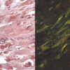

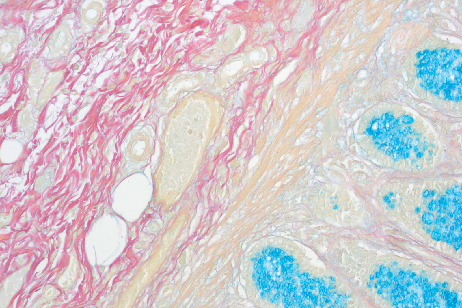
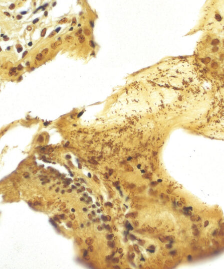
The Colloidal Iron kit is used for visualisation of carboxylated and sulphated groups of acid mucins and proteoglycans. This method is based on the principle of binding positively charged ferric ions (Fe3+) to negatively charged endings of acid mucopolysaccharides and proteoglycans. Excessive reagents are rinsed while the bound ferric ions get visualised using the Prussian Blue reaction. In this reaction potassium ferrocyanide causes light blue precipitations of iron ferrocyanide to appear. Finally, the sections are exposed to Van Gieson stain that selectively stains different tissue structures and in turn creates clear and visually rich contrast. The method can be combined with the PAS method; that way glycogen and neutral mucopolysaccharides would get differentially stained characteristically magenta.
Colloidal Iron kit
Six-reagent kit used for visualisation of carboxylated and sulphated groups of acid mucopolysaccharides and proteoglycans. This method can be combined with the PAS method; that way glycogen and neutral mucopolysaccharides would get differentially stained characteristically magenta.
Description
Additional information
| Size | |
|---|---|
| Brand | |
| Stain pack | |
| Stain Category | Carbohydrates |
Downloads
Related products
Stain Kits
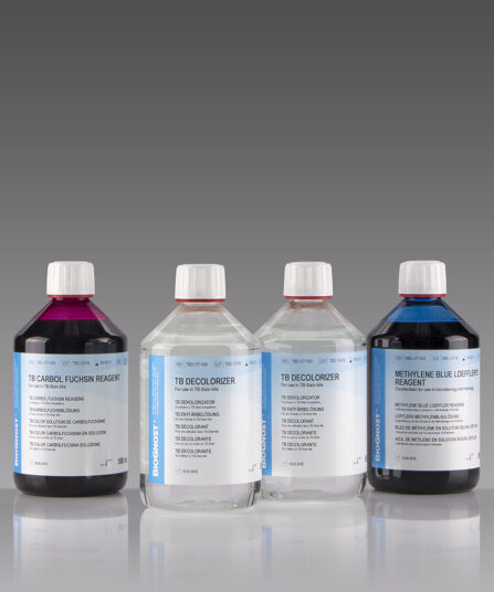
TB-Stain Hot Kit
Three-reagent kit for staining acid-fast bacteria. Contains TB Carbol Fuchsin reagent, double amount of TB Decolorizer and Methylene Blue Loeffler’s reagent as counterstain.
4 x 100ml bottles.

Stain Kits
Bio-Diff kit 3 x 500ml
Three-reagent kit that contains fi xative agent, red and blue components for fast and effective staining. Each kit contains buffer tablets for consistent staining results.

Stain Kits
TB-Stain Histo kit
Three-reagent kit for staining acid-fast bacteria (pathogenic mycobacteria) in histology sections, sputum, smears and culture smears according to Ziehl-Neelsen. Heating of the carbol-fuchsin solution is avoided in this protocol hence omitting the release of hazardous phenolic vapours.
Stain Kits
Warthin Starry kit
Five-reagent kit for staining Spirochaeta, Helicobacter pylori, Microsporidia and Legionella pneumophila. The kit contains 12 jars with gelatine that enables both incubation and staining of sections, as well as other reagents that enable precipitation of silver on the bacterial surface. The bacteria are found in the mucus of the surface epithelium, in the apical gastric glands and in the gastric mucosa.
HE Rapid Staining kit- frozen and paraffin sections
Ready-to-use eight-reagent kit (in 16 containers that can be used as staining jars) for rapid HE staining of frozen and paraffin tissue sections in histopathology. Contains xylene substitute as clearing agent and xylene substitute-based medium for permanent section covering.
For 100 tests.
Stain Kits
Gomori Trichrome kit
Five-reagent kit for staining muscle, collagen fibre and nuclei, contains blue counterstain. The kit can be used to contrast skeletal, cardiac or smooth muscle.
Stain Kits
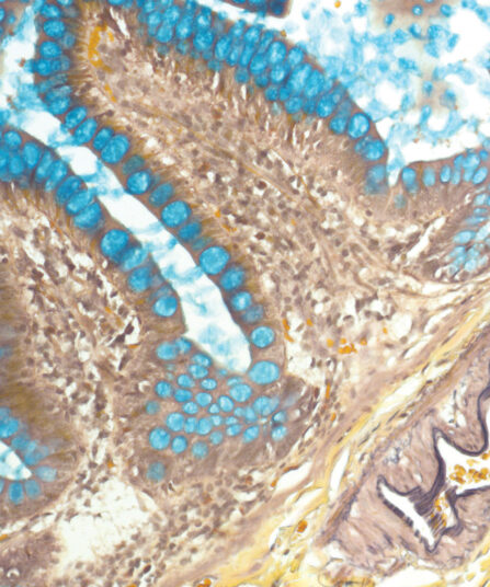
Alcian Yellow Toluidine Blue kit
Six-reagent kit for staining Helicobacter pylori in gastric tissue sections. This method is one of the most popular non-silver methods for staining of H. pylori, where bacteria are stained blue in contrast to yellow mucins.