HISTOLOGY
The word ‘Histos’ is Greek for web or tissue and ‘logia’ is Greek for branch of learning, therefore ‘Histology’ means the study of the microscopic structure of tissues.
More...
Tissue was first used to describe the different textures of body parts being dissected by an anatomist. Histology first came into use in the 1700s, but it is thought it began in Italy in the 1600s when scientist Marcello Malpighi experimented with insects, botany, and embryology.
There are four basic tissue types including Epithelium, Connective Tissue, Nervous Tissue, and Muscle. Each type contains subtypes that may look different but share similar characteristics. Epithelium tissue is the thin tissue forming the outer layer of a body’s surface including skin and internal hollow structures
Body tissues grow by increasing the number of cells that make them up. Cells in many tissues in the body divide and grow very quickly until we become adults
Our bodies are made up of about a hundred million million (100,000,000,000,000) tiny cells. You can only see them under a microscope
Cells group themselves together to make up the tissues and organs of our bodies. They are a bit like building blocks. The diagram below shows what cells look like when they are grouped together
show less
Stain Kits
A.F.O.G. kit
Six-reagent Acid Fuchsin Orange G kit for selective staining of glomerular protein deposits and collagen in kidney biopsies. Nuclear stain is obtained with Weigert ferric hematoxylin, cytoplasm with Orange G and highly selective collagen stain with Aniline blue.
Stain Kits
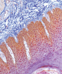
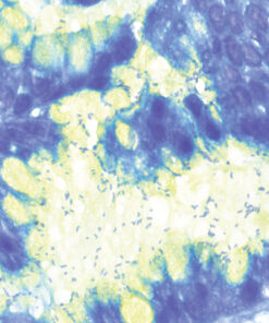
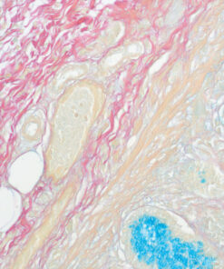
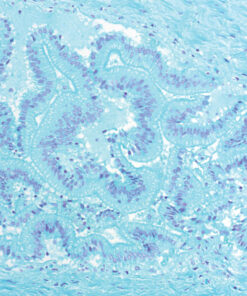
Alcian Blue – P.A.S. kit
Seven-reagent Alcian Blue – Periodic Acid-Schiff kit for staining acid mucopolysaccharides according to Mowry. Enables differentiation between acid mucins (stained light blue) and neutral mucins, glycogens and glycoproteins (stained red, magenta).
Stain Kits
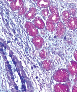
Alcian Blue pH 1.0 kit
Three-reagent kit for staining heavily sulfated mucopolysaccharides. Slides are counterstained with Nuclear Fast Red (Kernechtrot) reagent to fully detect the presence of Alcian Blue positive staining.
Stain Kits
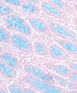
Alcian Blue pH 2.5 kit
Three-reagent kit for staining acid mucopolysaccharides according to Dorling and sulphated and carboxylated sialomucins blue. Slides are counterstained with Nuclear Fast Red (Kernechtrot) reagent to fully detect the presence of Alcian Blue positive staining.
Stain Kits
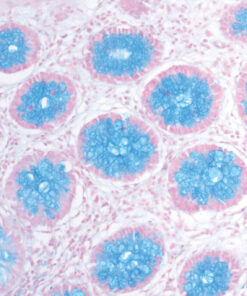
Alcian Yellow Toluidine Blue kit
Six-reagent kit for staining Helicobacter pylori in gastric tissue sections. This method is one of the most popular non-silver methods for staining of H. pylori, where bacteria are stained blue in contrast to yellow mucins.
Stain Kits
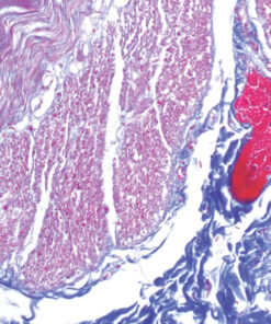
Azan Trichrome kit
Five-reagent kit for connective tissue staining according to Mallory. Used for visualisation of muscle fibres, collagen, glial cells, glomerular cells and erythrocytes.
Stain Kits
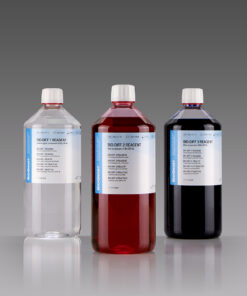
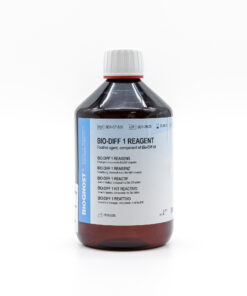
Bio-Diff Kit
Three-reagent kit that contains fixative agent, red and blue components for fast and effective staining. Each kit contains buffer tablets for consistent staining results.
3×100 ml bottles
Stain Kits
Bio-Diff RTU kit
Ready-to-use three-reagent kit with reagents stored in containers that can be used as staining jars. Kit contains fixative agent, red and blue components for fast and effective staining and buffer tablet for consistent staining results.
Stain Kits
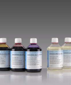
BioGram 4 kit
Four-reagent kit for identification of bacteria according to Gram. Kit contains Gram Crystal Violet 1% solution, stabilized Gram Lugol solution, double amount of Gram Decolorizer solution 2 and Gram Safranin solution as counterstain.
5×100 ml bottles
Stain Kits
BioGram ECO kit
Four-reagent phenol-free kit for the identification of bacteria according to Gram. Kit contains Gram Crystal violet, phenol free reagent, Gram Sodium hydrogencarbon, solution, stabilized Gram Lugol solution, double amount of Gram Decolorizer solution 2 and Gram Safranin solution as counterstain.
2×50 mL+4×100 mL bottles
Stain Kits
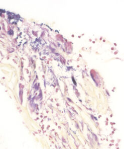
BioGram Histo kit
Five-reagent kit for identification of bacteria according to Gram. For differentiation between Gram-positive and Gram-negative bacteria in histology sections.
Stain Kits
Colloidal Iron kit
Six-reagent kit used for visualisation of carboxylated and sulphated groups of acid mucopolysaccharides and proteoglycans. This method can be combined with the PAS method; that way glycogen and neutral mucopolysaccharides would get differentially stained characteristically magenta.
Stain Kits
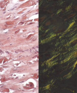
Congo Red Highman kit
Three-reagent kit for staining amyloids, characteristic for use of alkaline solution as differentiation medium in order to avoid undesirable non-specific colouration of cellular substances. Amyloid deposits display green colouration under polarised light.

Stain Kits
Congo Red Puchtler kit
Three-reagent kit for staining amyloids, characteristic by its high ionic strength and pH enhancing the specificity of Congo Red dye binding to amyloid clusters. This method developed by Puchtler remains the gold standard for amyloids in tissue sections. Amyloid clusters have the property of double refraction that enables green colouration under polarised light.
Stain Kits
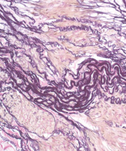
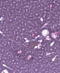
Elastica-Van Gieson kit
Four-reagent kit for staining elastic fibres and differentiation between elastic tissue, collagen and other types of connective tissue. The rapid method enables a satisfactory result with shorter section staining time.
Stain Kits
Eosin and Nigrosin Vital kit
Two-reagent kit containing separate dyes for rapid detection of sperm vitality and simple visualisation of dead and living sperm cells. The Nigrosin stain provides dark background for easier recognition of both viable and non-viable spermatozoa.
Stain Kits
Eosin-Nigrosin Vital
Fast detection (one-step detection) of sperm vitality and visualisation of dead and living sperm cells with one reagent. A simple, easy and fast method for semen analysis.
Stain Kits
Feulgen kit
Five-reagent DNA staining kit according to Feulgen. For use in semiquantitative DNA determination in histological and cytological samples. The specimen is first treated with hydrochloric acid creating an aldehyde group of DNA that can be visualised by Schiff (BioSchiff) reagent. This reaction is specific for nuclear DNA.
Stain Kits
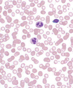
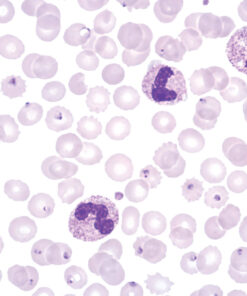
Field kit
Ready to use two-reagent kit for rapid and efficient staining and detection of parasites in haematology samples. Primarily used for staining thin and thick blood smears (dense drop) for purpose of diagnosing blood parasites. Reagents are stored in containers that can be used as staining jars.
Stain Kits
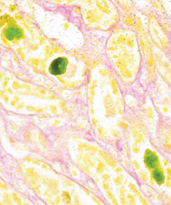
Fouchet-Van Gieson kit
Three-reagent kit for visualisation of bilirubin and collagen according to Kutllick. Bilirubin is a yellow-brown pigment, but changes to green due to oxidation induced by Fouchet reaction. Green bilirubin can easily be detected on yellowish and pink coloured background.